ABCDEF Model
A · Axis
Lead I QRS polarity
Choose the predominant QRS direction
Lead aVF QRS polarity
Used with lead I to determine the frontal axis quadrant
Lead aVR QRS polarity
A dominant positive QRS supports a northwest axis pattern
Select lead I, aVF, and aVR polarity.
Baseline Axis Change (Optional)
Machine Calculated QRS Axis:
°
°
Choose +/− and enter both axes to compare.
ABCDEF Assessment
AAxis
Northwest axis with lead I negative and aVF negative
BBroad
QRS duration greater than 200 ms
ms
CConcordance
All QRS complexes in V1 through V6 predominantly positive or all predominantly negative
DDissociation
AV dissociation, capture beats, or fusion beats
EEarly
Slow initial QRS activation or time to peak greater than 40 ms
FFragmentation
Josephson sign with notching near the nadir of the S wave in V1
Simplified Approach: ABCDEF Model - My preferred approach
A: Axis
- Is there northwest axis (-90 to -180 degrees)?
- 90% specific for VT
- aVR is POSITIVE and Lead I and aVF are NEGATIVE


B: Broad Complexes
- QRS duration >200ms is 85-90% specific for VT
- Use the widest clearly measurable QRS complex (typically V1 or V2)

C: Concordance
- Are the precordial leads entirely negative or entirely positive (aka no rS complexes seen)
- 97% specific for VT

D: Dissociation
- Is there p wave dissociation?
- Are there fusion or capture beats?


E: Early QRS Slurring
- Is the early part of the QRS depolarization fast or slow
- If slow = VT
- Can either use time to peak of R wave >40ms or examine the morphology to see if there is slurring of the initial depolarization

F: Fragmention of S wave in V1
- A notched S wave in V1 (also known as Josephson sign)
- 97-100% specific for VT
- Also known as Josephson sign

Complex Model: Basel, Brugada, Verecki
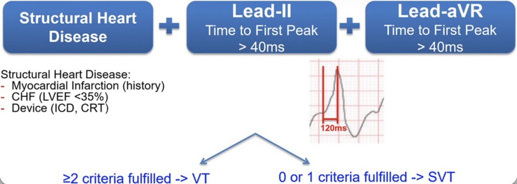
Basel + Brugada + Vereckei Criteria
Basel Criteria
1. Clinical high-risk feature for VT present?
Examples prior MI, structural heart disease, prior VT, severe cardiomyopathy
2. Lead II time to first peak > 40 ms?
3. aVR time to first peak > 40 ms?
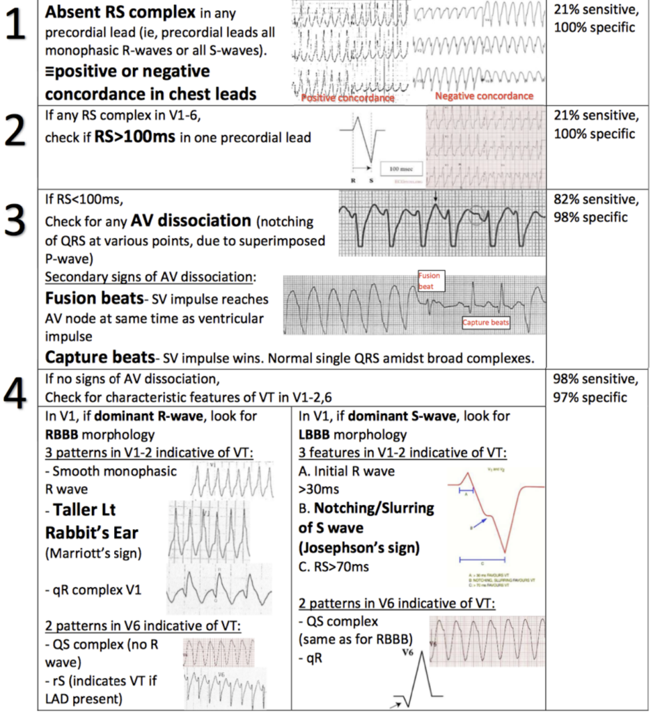
Brugada Criteria
1. No RS complex in all precordial leads?
2. RS interval > 100 ms in any precordial lead?
3. AV dissociation present?
4. Morphology criteria for VT met?
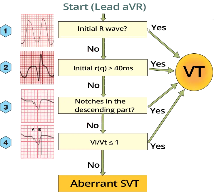
Vereckei Criteria
1. Initial R wave in aVR?
2. Initial r or q wave in aVR ≥ 40 ms?
3. Notching on the downstroke of a predominantly negative QRS in aVR?
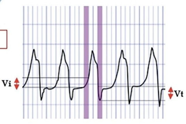
4. Vi/Vt ≤ 1?
Initial ventricular activation slower than terminal portion
Basel Algorithm
- 2022 multicenter study proposing the Basel algorithm to differentiate VT from SVT with aberrancy in a wide-complex tachycardia
- VT diagnosed if at least 2 of 3 criteria are present
- High-risk (MI, EF<35%, ICD/CRT, other high risk structural disease)
- Lead II time to peak >40ms
- Lead aVR time to peak >40ms
- In derivation and validation cohorts the algorithm showed ~92–93% sensitivity and ~89–90% specificity for VT
- Diagnostic accuracy comparable to Brugada and Vereckei algorithms but significantly faster to apply in usability testing
Brugada Criteria
If any are + then assume VT
- Absence of RS complex in all precordial leads (V1–V6)
- If every QRS is either monophasic R or QS → VT
- RS interval >100 ms in any precordial lead
- Measure from R onset to S nadir
- AV dissociation present
- P waves independent of QRS
- Morphologic VT criteria
- Evaluate QRS morphology in V1/V2 and V6 for VT patterns
Vereckei (aVR) Algorithm
If any are + then assume VT
- Initial R wave in aVR
- Initial r or q wave in aVR ≥40 ms
- Notching on the downstroke of a predominantly negative QRS in aVR
- Vi/Vt ≤1 (initial ventricular activation slower than terminal portion)
References
- Moccetti, F., Yadava, M., Latifi, Y., Strebel, I., Pavlovic, N., Knecht, S., Asatryan, B., Schaer, B., Kühne, M., & Henrikson, C. A. (2022). Simplified integrated clinical and electrocardiographic algorithm for differentiation of wide QRS complex tachycardia: The Basel algorithm. JACC: Clinical Electrophysiology, 8(7), 831–839. https://doi.org/10.1016/j.jacep.2022.03.017
- Brugada, P., Brugada, J., Mont, L., Smeets, J., & Andries, E. W. (1991). A new approach to the differential diagnosis of a regular tachycardia with a wide QRS complex. Circulation, 83(5), 1649–1659. https://doi.org/10.1161/01.CIR.83.5.1649
- Vereckei, A., Duray, G., Szénási, G., Altemose, G. T., & Miller, J. M. (2008). New algorithm using only lead aVR for differential diagnosis of wide QRS complex tachycardia. Heart Rhythm, 5(1), 89–98. https://doi.org/10.1016/j.hrthm.2007.09.020
- Vereckei, A., Duray, G., Szénási, G., Altemose, G. T., & Miller, J. M. (2007). Application of a new algorithm in the differential diagnosis of wide QRS complex tachycardia. European Heart Journal, 28(5), 589–600. https://doi.org/10.1093/eurheartj/ehl473
- Katritsis, D. G., & Brugada, J. (2020). Differential diagnosis of wide QRS tachycardias. Arrhythmia & Electrophysiology Review, 9(3), 155–160. https://doi.org/10.15420/aer.2020.20
-
Buttner, R. (2026, July 17). VT versus SVT: It’s as easy as ABCDE.
Life in the Fast Lane.
VT versus SVT: It’s as easy as ABCDE