QTc in Conduction Disease (JT Interval)

JT Interval Calculator

Assess QT interval when the QRS is prolonged. Hodges method preferred.

Measurements

QT interval
Measured QT interval
ms
QRS duration
JT is most useful when QRS ≥120 ms
ms
Heart rate
bpm
Correction method
Hodges: JTc = JT + 1.75 × (HR − 60)

Why Use JT Instead of QT?


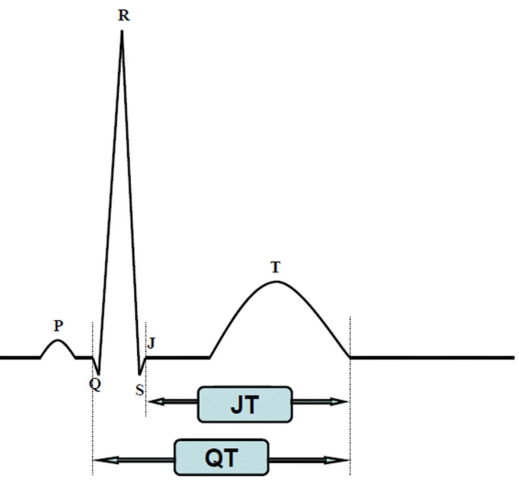
The JT interval more directly reflects ventricular repolarization. It begins at the end of the QRS complex and ends at the end of the T wave.

When the QRS is prolonged, the QT interval also becomes longer because QT includes both ventricular depolarization and repolarization. QTc may therefore overestimate repolarization abnormality in a wide-QRS rhythm. The ACC specifically endorses use of JTc as a validated approach to analyze QT interval in conduction disease.

  • Left bundle branch block
  • Right bundle branch block
  • Ventricular pacing
  • Pre-excitation
  • Nonspecific intraventricular conduction delay
  • Ventricular escape rhythms
QT and JT interval electrocardiogram diagram

Calculation


JT interval
JT = QT − QRS
Hodges correction
JTc = JT + 1.75 × (HR − 60)
Framingham correction
JTc = JT + 154 × (1 − RR)
Fridericia correction
JTc = JT ÷ RR1/3

JTc Interpretation


JTc Interpretation Clinical Meaning
<360 ms Below common high threshold Not prolonged by the commonly used 360 ms screening threshold.
≥360 ms Prolonged Commonly used threshold for abnormal JTc in wide-QRS assessment.
JTc reference limits vary by correction method and population. There is no single universally accepted cutoff.

Key Evidence


Zulqarnain et al. 2015

Risk of Mortality Associated With QT and JT Intervals at Different Levels of QRS Duration (NHANES III)
Population
8,025 adults
Deaths
3,045
Wide QRS
≥120 ms

In participants with QRS ≥120 ms, prolonged JT was substantially more strongly associated with mortality than prolonged QT after multivariable adjustment.

Adjusted hazard ratio for all-cause mortality with QRS ≥120 ms. “Prolonged” was defined as >95th percentile: QT >462 ms and JT >362 ms; these were uncorrected intervals, not JTc cutoffs.

Tsai et al. 2014

QTc Compared With JTc for Monitoring Drug-Induced Repolarization Changes During Ventricular Pacing
Population
22 patients
Drugs
Sotalol / dofetilide
Comparison
Native vs paced

This study directly tested the clinical problem that makes JT useful. When a patient is ventricularly paced, the paced QRS becomes wide and lengthens the QT even if repolarization itself has not changed proportionally. During sotalol or dofetilide loading, the mean increase in peak QTc was therefore smaller during ventricular pacing than during native conduction (7% vs 12%, P=.003). In contrast, peak JTc was essentially unchanged between native conduction and ventricular pacing (383±37 vs 386±32 ms, P=.66).

Why this matters: QTc can become misleading when pacing widens the QRS. JTc removes the depolarization component and more faithfully follows drug-induced repolarization change, making it particularly useful when monitoring QT-prolonging antiarrhythmics in ventricular-paced rhythms.
Mean increase in peak QTc during antiarrhythmic loading. Native conduction showed a 12% increase versus 7% during ventricular pacing (P=.003), while peak JTc did not differ significantly between conduction states.

Correction Method Evidence


Rautaharju 2004

QTc − QRS is not ideal

In 11,739 adults with normal conduction and 1,251 with major ventricular conduction defects, simple QTc − QRS retained substantial residual heart-rate dependence. Rate-adjusted JT or a model accounting for both heart rate and QRS duration performed better.

Chiladakis 2012

Hodges performs well in BBB

Hodges showed the least heart-rate dependent variability of QTc and JTc across heart rates in patients with bundle branch block. Framingham and the Nomogram method also performed well, while Bazett exaggerated heart-rate dependence.

Funk 2021

Wide-QRS QT requires adjustment

A review focused on QT assessment in ventricular conduction delay and ventricular pacing recommended either the Bogossian approach or use of the JT interval followed by Hodges or Framingham heart-rate correction.

Practical Point

Avoid Bazett when possible

Bazett correction becomes increasingly distorted at heart rates farther from 60 bpm and performed poorly in comparative assessment of wide-QRS rhythms.

References
1
Zulqarnain MA, Qureshi WT, O’Neal WT, Shah AJ, Soliman EZ. Risk of mortality associated with QT and JT intervals at different levels of QRS duration. Am J Cardiol. 2015;116(1):74-78.
View study
Mortality / Prognosis
2
Rautaharju PM, Zhang ZM, Prineas R, Heiss G. Assessment of prolonged QT and JT intervals in ventricular conduction defects. Am J Cardiol. 2004;93(8):1017-1021.
View study
Conduction Defects
3
Chiladakis J, Kalogeropoulos A, Koutsogiannis N, Zagkli F, Vlassopoulou M, Chouchoulis K, Alexopoulos D. Optimal QT/JT interval assessment in patients with complete bundle branch block. Ann Noninvasive Electrocardiol. 2012;17(3):268-276.
View study
Bundle Branch Block
4
Tsai SF, Houmsse M, Dakhil B, Augostini R, Hummel JD, Kalbfleisch SJ, et al. QTc compared to JTc for monitoring of drug-induced repolarization changes in the setting of ventricular pacing. Heart Rhythm. 2014;11(3):485-491.
View study
Ventricular Pacing
5
Funk MC, Cates KW, Rajagopalan A, Lane CE, Lou J. Assessment of QTc and risk of torsades de pointes in ventricular conduction delay and pacing: a review of the literature and call to action. J Acad Consult Liaison Psychiatry. 2021;62(6):603-613.
View review
Drug Monitoring
6
Chiladakis J, Kalogeropoulos A, Zagkli F, Koutsogiannis N, Chouchoulis K, Alexopoulos D. Predicting torsade de pointes in acquired long QT syndrome: optimal identification of critical QT interval prolongation. Cardiology. 2012;122(1):3-11.
View study
Torsades Risk
7
Rautaharju PM, Surawicz B, Gettes LS, et al. AHA/ACCF/HRS recommendations for the standardization and interpretation of the electrocardiogram: part IV: the ST segment, T and U waves, and the QT interval. J Am Coll Cardiol. 2009;53(11):982-991.
View guideline
AHA ECG Standards