Pulmonary Hypertension Severity
TTE Hemodynamics
Estimate RAP from IVC + hepatic vein Doppler
Add optional RV function markers
Right Heart Catheterization
Add right-heart reserve / PAH risk markers
Initial Workup
- Confirm the phenotype on TTE: TR velocity/RVSP, RV size and function, RA/IVC, septal shape, RVOT Doppler, left-sided filling pressures and valve disease.
- Labs: CBC, CMP/LFTs, TSH, BNP or NT-proBNP, HIV, ANA/CTD testing when clinically indicated.
- V/Q scan: screen for chronic thromboembolic pulmonary disease when PH is unexplained or a precapillary phenotype is suspected.
- PFTs + DLCO, oximetry/ABG, chest CT: define Group 3 lung disease/hypoxia and look for a pulmonary vascular phenotype disproportionate to parenchymal disease.
- Sleep-disordered breathing: test when clinically suspected, especially with obesity, nocturnal hypoxemia, or chronic hypercapnia.
- RHC: required to definitively diagnose and classify PH before PAH-specific therapy.
PH Classification
| Group | Mechanism | Common causes | Echo clue | Key next step |
|---|---|---|---|---|
| 1 | PAH / precapillary | Idiopathic/heritable, CTD, portal HTN, HIV, congenital heart disease, drugs/toxins | Small/normal left heart, RV pressure overload, short RVOT AccT/notching, RV/RA enlargement, abnormal RV-PA coupling | RHC + PAH workup; vasoreactivity testing only in appropriate idiopathic/heritable/drug-associated PAH |
| 2 | Left heart disease / postcapillary | HFpEF, HFrEF, cardiomyopathy, mitral/aortic valve disease | LA enlargement, LVH or LV dilation, grade ≥2 diastolic dysfunction, elevated E/e′, left-sided valve disease | Treat left-heart disease; use RHC when phenotype is uncertain or appears disproportionate |
| 3 | Lung disease / hypoxia | COPD/emphysema, ILD, CPFE, hypoventilation syndromes | PH may be present with lung disease; marked RV pressure overload out of proportion to lung disease should prompt a broader workup | PFT/DLCO, oxygenation, chest CT; optimize lung disease and hypoxemia |
| 4 | Pulmonary artery obstruction | CTEPH / chronic thromboembolic pulmonary disease, other PA obstruction | Often a precapillary RV pressure-load phenotype; echo cannot exclude CTEPH | V/Q scan, then CTEPH-center imaging/RHC if abnormal |
| 5 | Unclear / multifactorial | Sarcoid, hematologic disorders, CKD, complex systemic disease | Variable and often mixed | Mechanism-specific workup and specialty input |
RHC defines pulmonary hypertension, but the pressure itself is only part of the story. A useful Mayo Clinic pearl to think about it is that PAP is the single glucose value and the right heart is the A1c. The pressure can fluctuate, and in advanced disease it can even fall as the RV loses the ability to generate pressure.
Pressure Quantification
| Grade | TRV | RVSP | mPAP on RHC |
|---|---|---|---|
| Normal | <2.8 m/s | ≤34 mmHg | ≤20 mmHg |
| Mildly elevated | 2.8-3.1 m/s | 35-49 mmHg | 21-30 mmHg† |
| Moderately elevated | 3.2-3.5 m/s | 50-69 mmHg | 31-40 mmHg† |
| Severely / markedly elevated | ≥3.6 m/s | ≥70 mmHg | >40 mmHg† |
RVSP equals sPAP only when there is no RVOT obstruction.
TTE Measurements
- TR peak velocity: interrogate from multiple windows and use the highest dense envelope. Average at least 3 beats in sinus rhythm and more beats with an irregular rhythm.
- Measure the chin, not the beard: trace the dense primary edge of the CW signal, not faint overgained signal.
- Incomplete signal: agitated saline, blood-saline contrast, or an ultrasound-enhancing agent can improve the envelope. Do not simply turn the gain up.
- RVSP: 4 × (TR peak velocity)² + RAP.
- sPAP: RVSP = sPAP only if there is no RVOT obstruction, pulmonic stenosis, or proximal PA stenosis.
- Severe TR: a dagger/triangular, truncated CW signal can underestimate RVSP from early RA-RV pressure equalization. A parabolic contour is more reliable.
- PA end-diastolic pressure: 4 × (PR end-diastolic velocity)² + RAP.
- Estimated mPAP: 0.61 × sPAP + 2 mmHg.
- If PAEDP is available: mPAP ≈ ⅓(sPAP) + ⅔(PAEDP).
- TRV threshold: ≥2.9 m/s supports PH. A TRV of 2.8 m/s can also support PH when at least 2 additional echo signs are present.
Right Atrial Pressure: IVC + Hepatic Vein Doppler
How to Actually Define Severity
Do not grade the patient by RVSP alone. The RV response to afterload is more clinically important. As PH progresses the RV enlarges, contractile reserve falls, the septum shifts leftward, LV filling falls, RAP rises, and eventually the RV may be unable to generate a very high sPAP.
RV size / remodeling
Use an RV-focused view. Linear dimensions help, but 3D RV volumes are better when available. Worsening RV enlargement, RV/LV ratio >1, septal flattening, and a progressively small/underfilled LV indicate maladaptation.
RV systolic function
TAPSE and S′ are useful but limited. FAC adds radial shortening. RV free-wall strain is more sensitive to early dysfunction. Mayo board pearl: strain becoming less negative than about -25% is concerning.
Afterload
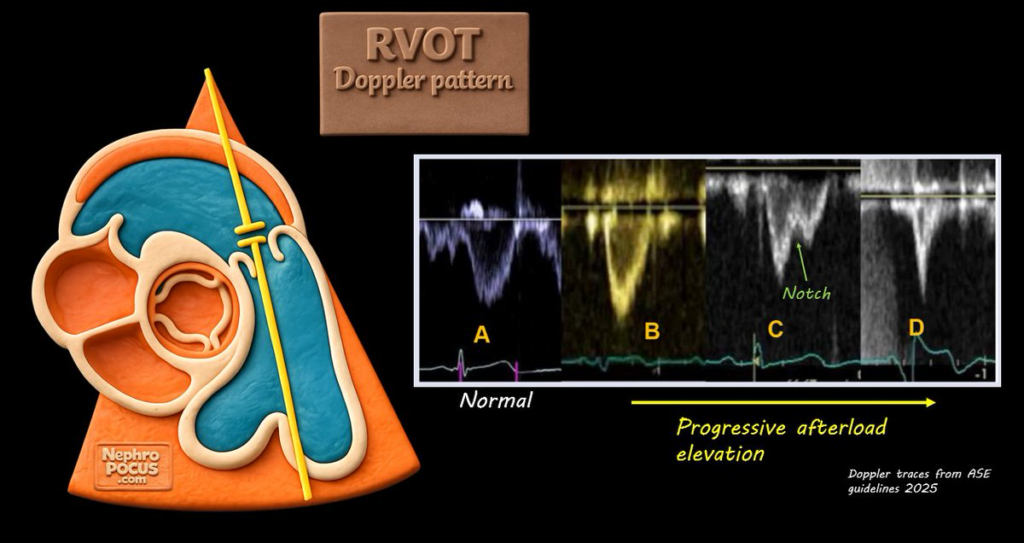
RVOT Doppler becomes more triangular with an early peak, shorter acceleration time, and midsystolic notch/W pattern as pulmonary vascular impedance increases. Pure postcapillary PH is less likely to show a classic high-PVR notch pattern.
RV-PA coupling
TAPSE/sPAP asks whether the RV is generating enough contraction for its afterload. Values around 0.3-0.4 mm/mmHg are associated with RV-PA uncoupling and worse outcomes.
Right-sided filling pressure
Rising RAP, RA enlargement, dilated IVC, loss of hepatic systolic predominance, and systemic venous congestion reflect a failing RV that is no longer compensating for the afterload.
Pericardial effusion
In PAH, a new effusion is a prognostic marker of chronic right-sided pressure burden. Tamponade physiology can be atypical when baseline right-sided pressures are very high.
RV Quantification Reference
| Measure | Normal | Mild | Moderate | Severe |
|---|---|---|---|---|
| TAPSE | >1.7 cm | 1.3-1.7 | >1.0-<1.3 | ≤1.0 |
| S′ | >9.5 cm/s | 7.2-9.5 | >5.0-<7.2 | ≤5.0 |
| FAC | >35% | 30-35% | 23-29% | ≤22% |
| RV free-wall strain |%| | >20% | 15-20% | 11-<15% | <11% |
| RV basal diameter | <4.1 cm | 4.1-4.4 | >4.4-4.9 | >4.9 |
| RA area | <19 cm² | 19-22 | >22-24 | >24 |
| RVOT AccT | >105 ms | 80-105 | 60-<80 | ≤60 |
RV Afterload
- Best quantified invasively by PVR, which reflects the resistive component of pulmonary vascular load
- PVR = (mPAP − PAWP) / CO
- Normal is roughly <2 WU
- >2 WU is abnormal and supports a precapillary component
- PVR = (mPAP − PAWP) / CO
- RVOT Doppler: Increasing pulmonary vascular load produces an earlier systolic peak, shorter acceleration time, and eventually midsystolic notching/W-pattern. Notching is particularly suggestive of increased pulmonary vascular impedance and a precapillary phenotype.
- RVOT acceleration time
- <105 ms: may support PH
RVOT Doppler: Pulmonary Vascular Load
Onset of ejection → peak velocity
Reflects abnormal pulmonary vascular wave reflection
Pre- vs Postcapillary PH by Echo
TTE cannot assign a final PH group, but it is often very useful for the first major split: does this look like left-heart disease, or does it look pre-capillary? Obvious HFrEF or severe left-sided valve disease is straightforward. HFpEF is the harder phenotype.
- Normal or small left-sided chambers
- RV/LV basal ratio >1, RV hypertrophy or pressure-loaded RV
- Septal flattening / LVEI >1.1
- RVOT acceleration time ≤105 ms and/or midsystolic notch
- TRV elevated with abnormal PR end-diastolic velocity
- TAPSE/sPAP reduced
- Dilated IVC, RA enlargement, abnormal hepatic vein Doppler as disease advances
- LV hypertrophy or LV dilation
- Reduced LVEF, or a convincing HFpEF phenotype
- LA enlargement
- Grade ≥2 LV diastolic dysfunction / elevated left-sided filling pressure markers
- Mitral or aortic valve disease sufficient to explain PH
- Right heart may remain relatively preserved until Group 2 disease is advanced
RHC Measurements
- PH: mPAP >20 mmHg at rest.
- Precapillary PH: mPAP >20, PAWP ≤15, PVR >2 WU.
- Isolated postcapillary PH: mPAP >20, PAWP >15, PVR ≤2 WU.
- Combined post- and precapillary PH: mPAP >20, PAWP >15, PVR >2 WU.
- PVR: (mPAP - PAWP) / CO.
- TPG: mPAP - PAWP. Useful as a pressure gradient, but PVR is the current hemodynamic discriminator for a precapillary component.
- DPG: dPAP - PAWP. Can be reported but is not used to define CpcPH in current criteria.
- PAPI: (sPAP - dPAP) / RAP. As RV failure progresses, PA pulse pressure often narrows while RAP rises.
| Pattern | mPAP | PAWP | PVR |
|---|---|---|---|
| No resting PH | ≤20 | Any | Any |
| Precapillary PH | >20 | ≤15 | >2 WU |
| Isolated postcapillary PH | >20 | >15 | ≤2 WU |
| Combined post- + precapillary PH | >20 | >15 | >2 WU |
Exercise and Follow-up
- PAP should rise with exercise. An isolated peak sPAP is not enough to diagnose exercise PH because age, flow, and athletic conditioning matter.
- Current invasive definition of exercise PH: mPAP/CO slope >3 mmHg/L/min between rest and exercise.
- An exercise sPAP around 50-55 mmHg is a useful mental reference, but healthy older adults and athletes can reach the 55-60 mmHg range. Think pressure-flow relationship and RV reserve, not a single universal cutoff.
- Follow-up: a higher sPAP after effective therapy is not automatically worsening. If RV contractility and forward flow improve, the RV may be able to generate more pressure. Follow RV function, RA/RAP, symptoms, biomarkers, exercise capacity, and hemodynamics together.
Group 1 PAH
PAH is a precapillary phenotype after excluding Group 2, Group 3, and Group 4 causes. Treatment is risk-based and usually managed through a pulmonary hypertension center.
- PDE-5 inhibitors: sildenafil 20 mg TID or tadalafil 40 mg daily are common oral pathway options.
- Endothelin receptor antagonists: macitentan, ambrisentan, or bosentan depending on the clinical setting.
- sGC stimulator: riociguat is an alternative pathway agent; do not combine with a PDE-5 inhibitor.
- Prostacyclin pathway: parenteral epoprostenol/treprostinil or other prostacyclin-pathway therapy for higher-risk disease.
- Vasoreactivity testing: for the appropriate idiopathic, heritable, or drug-associated PAH phenotype. High-dose CCB therapy is reserved for true responders.