
MR Severity Grading
| Parameter | Mild | Moderate | Severe |
|---|---|---|---|
| EROA (cm²) | <0.2 | 0.2–0.39 | ≥0.4 |
| Regurgitant Volume (mL) | <30 | 30–59 | ≥60 |
| Regurgitant Fraction (%) | <30% | 30–49% | ≥50% |
| Vena Contracta (cm) | <0.3 | 0.3–0.69 | ≥0.7 |
Primary (Organic) MR – leaflet/chordal abnormality
- Mitral valve prolapse
- Flail leaflet (chordal rupture)
- Rheumatic disease
- Endocarditis
- MitraClip/iatrogenic
Secondary (Functional) MR – annular or ventricular abnormality
- Ischemic MR (due to papillary muscle displacement after MI, usually inferior/posterior MI)
- Dilated cardiomyopathy (annular dilation, papillary displacement)
- Restrictive LV remodeling
Ischemic / Functional MR
- Typically central or posteromedial jets.
- Mechanism: LV dilation → papillary muscle displacement → tethering of leaflets → incomplete coaptation.
- Ischemic MR after inferior MI often eccentric (posterior pap muscle dysfunction causing anterior jet).
When is MR Eccentric?
- Eccentric jet (wall-hugging, Coandă effect):
- Mitral valve prolapse (classic cause)
- Flail leaflet
- Chordal rupture
- Papillary muscle rupture (acute MR)
- Central jet:
- Functional/ischemic MR
- Annular dilation
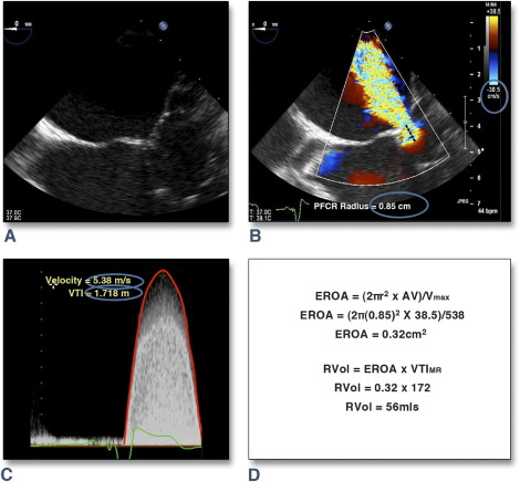
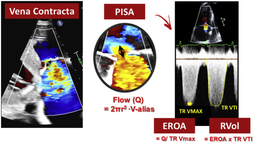
How to Measure
- EROA (PISA): Color Doppler → adjust Nyquist (~30–40 cm/s) → measure PISA radius (r) → Flow = 2πr² × aliasing vel → get peak MR Vmax (CW) → EROA = Flow ÷ Vmax
- RVol: RVol = EROA × MR VTI (trace CW jet)
- RF: RF = (RVol ÷ LV stroke vol) ×100% (LVSV via LVOT or volumes)
- VC: Parasternal long-axis, color Doppler, narrowest jet, avg early–mid systole.
Use ≥2–3 concordant findings; integrate with jet area, pulm vein flow, LV size.
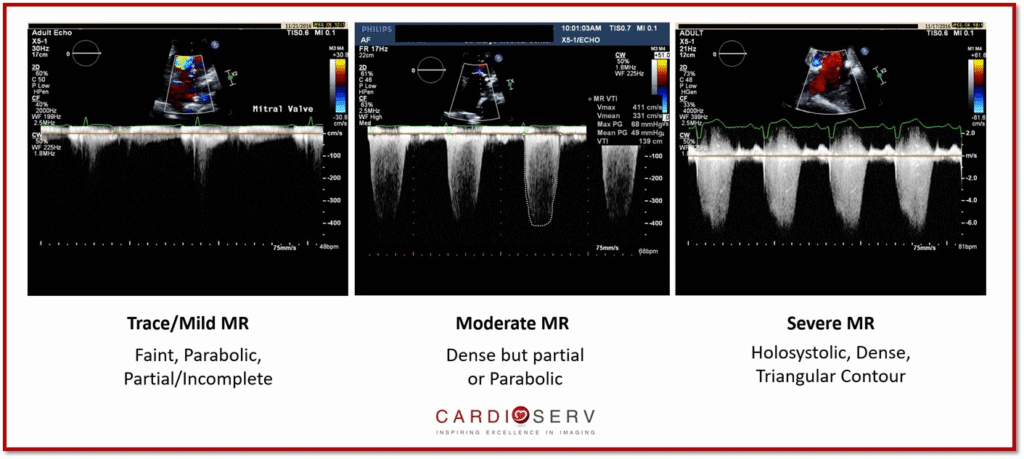
Qualitative Assessment (CW Doppler)
- Mild: Faint, parabolic, often <4m/s
- Moderate: Denser, parabolic, ~4m/s
- Severe: Very bright/dense, triangular (late-peaking), often 5-6m/s
Qualitative Assessment (Jet Length)
- Break the LA into thirds
- Depending on how far the jet travels backwards to the LA can indicate potential severity
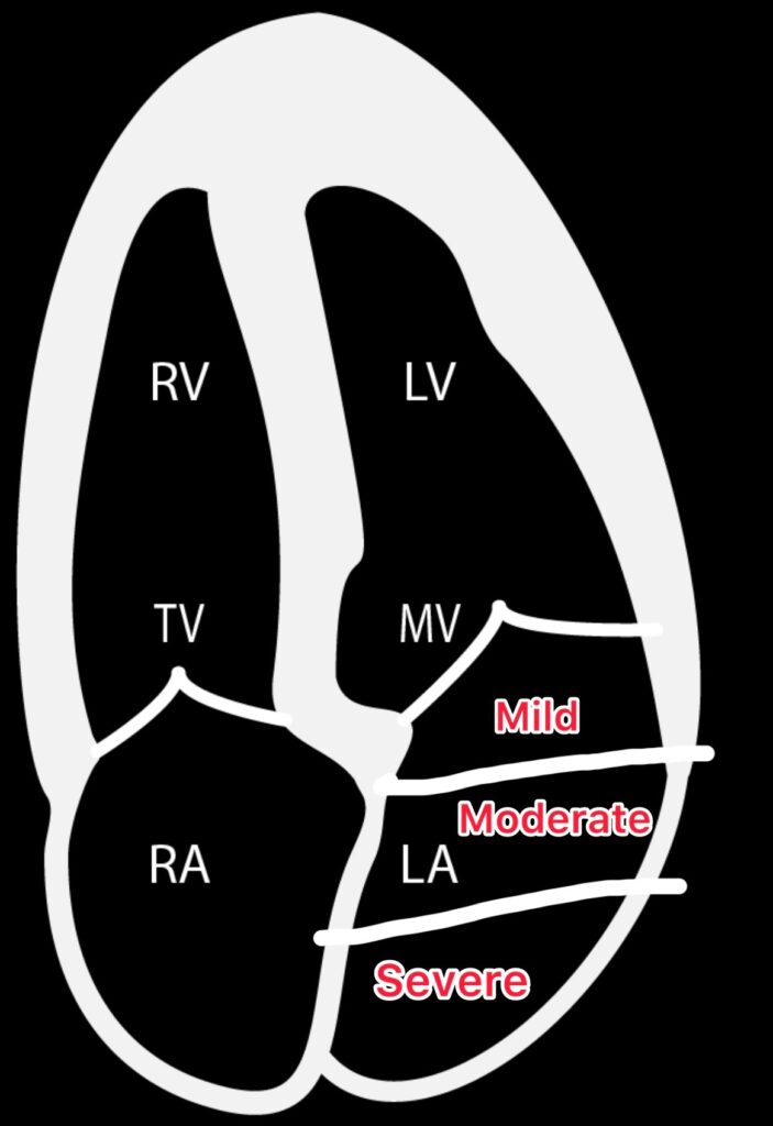
- A = Mild
- B = Moderate
- C = Severe
MR Severity Calculator
| Severity | EROA | RVol |
|---|---|---|
| Mild | <0.2 | <30 |
| Mild–Moderate | 0.2–0.29 | 30–44 |
| Moderate–Severe | 0.3–0.39 | 45–59 |
| Severe | ≥0.4 | ≥60 |