
Diastolic Function
Mitral Annular e′
Is there diastolic dysfunction?
Optional lateral e′
Mitral Inflow
What grade is it?
E/e′
Do they have elevated filling pressure?
TR Velocity
Is there PH?
Supportive LAP Markers
Optional. Select abnormal findings when primary markers are discordant.
Diastology Decision Tree
Reduced e′ velocity?
Septal ≤6 · lateral ≤7 · average ≤6.5 cm/s
Increased E/e′?
Septal ≥15 · lateral ≥13 · average ≥14
Increased TR velocity / PASP?
TR velocity ≥2.8 m/s · PASP ≥35 mmHg
E/A ≤0.8?
≥1 abnormal supportive marker?
E/A ≥2?
Primary Measurements
- Mitral annular tissue Doppler
- Mitral inflow Doppler
- E/e’
- Right ventricular systolic pressure
- Left atrial volume index
- Left atrial strain
- Pulmonary vein Doppler
Tissue Doppler- Do they have diastolic dysfunction?
Mitral annular motion reflects LV relaxation.
- Septal e’ velocity < 6 cm/s is abnormal
- Lateral e’ < 7 cm/s can also be used, but septal e’ is preferred and easier to measure
- If e’ is normal, the patient almost certainly has normal diastolic function and you are essentially done with diastology
- This is by far the most important measurement
Tissue Doppler Velocities
- s’ = LV systolic annular velocity
- e’ = early diastolic annular velocity, reflecting LV relaxation
- a’ = late diastolic annular velocity, reflecting atrial kick/late LV filling
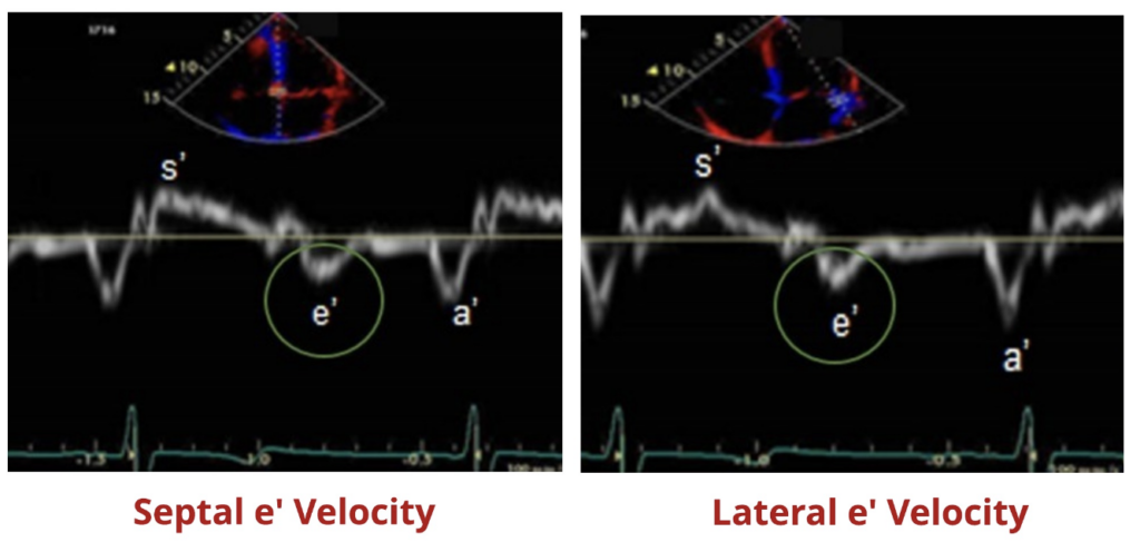
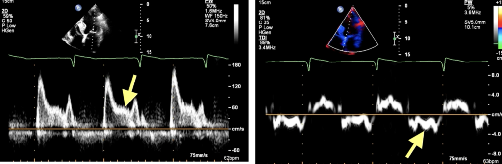
How to Measure Medial e’
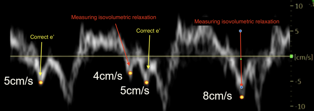
- Measure the bright modal envelope
- Avoid measuring artifact
- Measure the e’ envelope nearest to the a’ wave
- The preceding wave measured at 4 cm/s is isovolumetric relaxation and is not e’
- In this example, e’ < 6 cm/s, consistent with impaired relaxation
Mitral Inflow Doppler- How severe is their diastolic dysfunction?
- Measure using PW doppler at the mitral leaflet tips
- If e' is abnormal, mitral inflow velocity helps grade the severity of diastolic dysfunction
Normal
- E > A
- E/A > 0.8
- LV relaxes, pressure decreases and pressure rushes from high to low pressure area composing the majority of LV filling (E wave) with small component from atrial kick (A wave)
Grade I: Impaired Relaxation
- A > E
- E/A < 0.8
- Usually long deceleration time (> 240ms)
- LV is stiff so when MV opens, no longer low pressure in LV in early diastole so lower amplitude of E wave/slower deceleration time and more reliant on atrial kick to fill the LV but LAP normal
Grade II: Pseudonormal
- E > A
- E/A 0.8-2.0
- e’ is abnormal, so you know the apparently normal E/A pattern is actually abnormal
- LV is stiff but LAP is high enough to push blood out into the ventricle in early diastole
Grade III: Restrictive Filling
- E >> A
- E/A > 2.0
- Usually short deceleration time (< 160ms)
- LAP keeps climbing and get more rapid/early diastolic filling with very short deceleration time with very little atrial filling
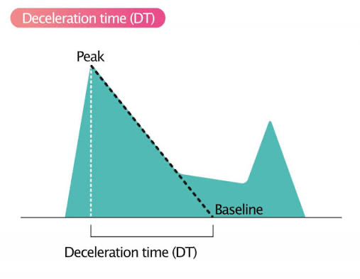
Deceleration time
- Measured from peak E wave to return to baseline
- Normal: 160-240ms
- Long DT > 240ms = impaired relaxation/grade I pattern
- Short DT < 160ms = restrictive filling / high LA pressure
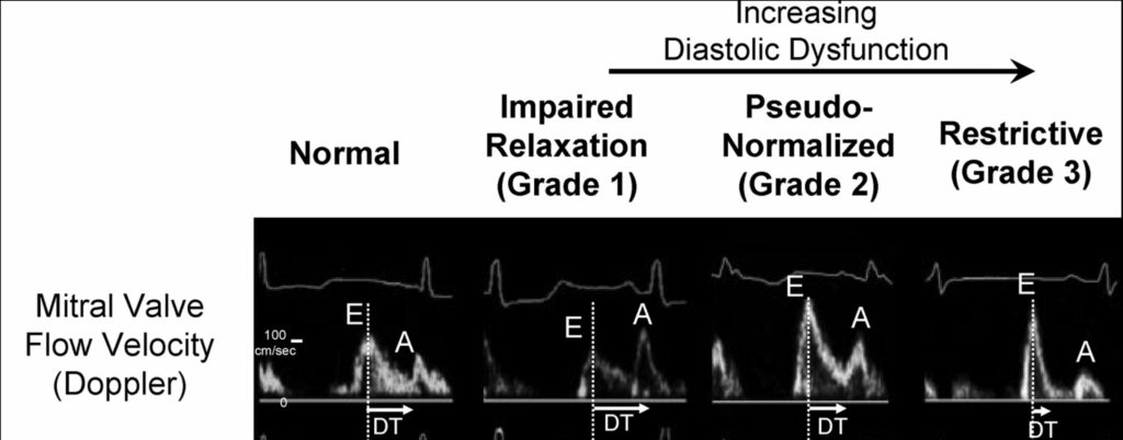
Mitral Inflow: Diastolic Dysfunction Pattern
normal
E/e’ Ratio- What is their filling pressure?
- E is taken from pulse wave Doppler mitral inflow
- e’ is taken from tissue Doppler of the mitral annulus
- E/e’ > 15 indicates elevated filling pressures/PCWP
- Specific but not sensitive
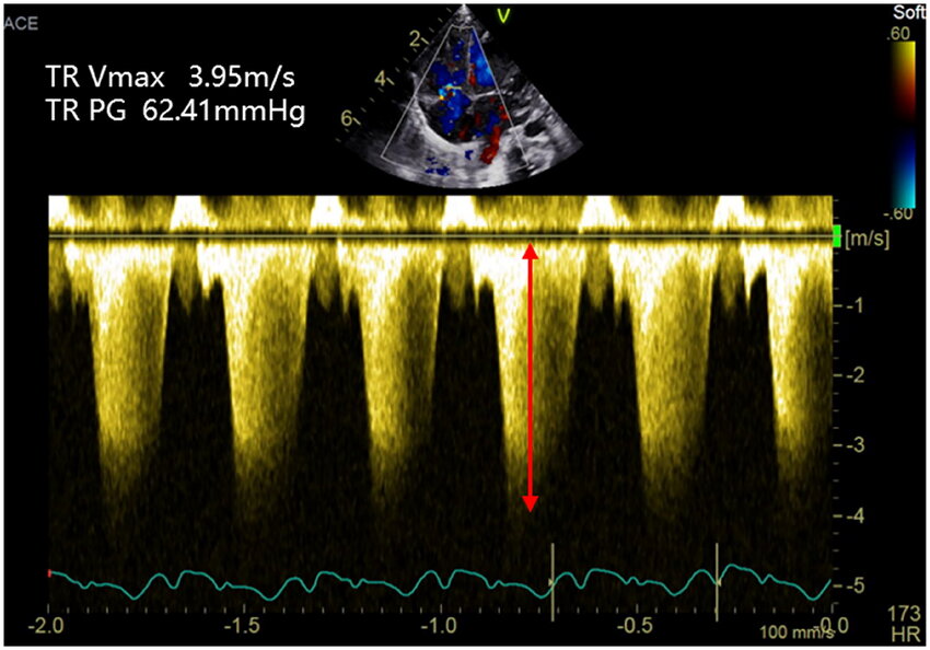
RVSP- Do they have pulmonary hypertension?
- Estimate RVSP using the TR peak velocity.
How to interpret
- TR peak velocity > 2.8 m/s is abnormal
- RVSP/sPAP > 25 mmHg is abnormal
- About two-thirds of patients with high LA pressure have pulmonary hypertension
- If they have pulmonary hypertension, it may be due to elevated LA pressure, though other confounders should be considered, such as COPD
Putting It All Together
Normal Diastolic Function
- e’ > 7 cm/s
- E/e’ < 15
- TR velocity < 2.8 m/s
Grade I Diastolic Dysfunction
- Medial e’ is abnormal, < 6 cm/s
- E/e’ is normal
- No pulmonary hypertension
- E < A
- Long deceleration time
Some patients fall between grade I and grade II. Diastology is often a spectrum.
Grade II/III Diastolic Dysfunction
- e’ < 6 cm/s
- E/e’ > 15
- TR velocity > 2.8 m/s
Then use mitral inflow to distinguish:
- Grade II: E/A < 2
- Grade III: E/A >> 2
What If It Is Not Clear?
Use additional markers of chronic pressure load and LA function.
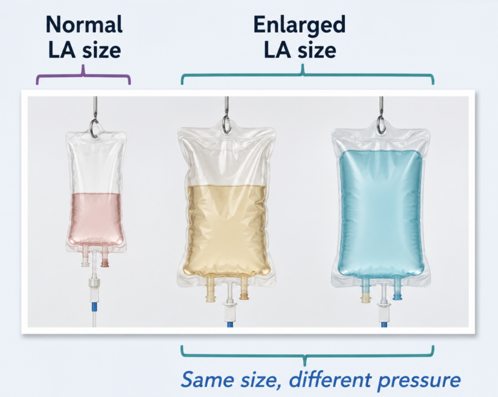
Left Atrial Enlargement
- Normal LA size: no evidence of long-term pressure elevation
- Enlarged LA: suggests chronic adverse pressure load
- LA enlargement alone does not tell you current filling pressures.
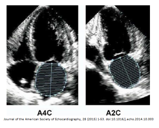
Left Atrial Volume Index
- LAVI > 34 mL/m² is abnormal
- Not a perfect measure
- If LAVI is around 50 mL/m², this is almost certainly abnormal
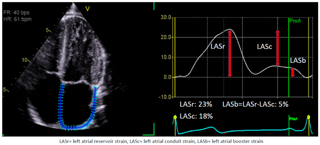
Left Atrial Strain
- Measure in the apical 4-chamber view
- Trace the LA in systole when the LA is largest
- Software generates the LA strain curve
- Left atrial strain looks at the contractility/function of the LA.
- Normal LA strain > 35%
- Clearly abnormal LA strain < 20%
Pulmonary Vein Doppler
Normal flow into the left atrium should be systolic predominant.
- S > D: likely normal LA pressure
- D > S: likely abnormal LA pressure
L Wave
- Extra wave in early/mid diastole that can indicate increased LV filling pressures
When not to try assessing diastology?
- Mitral prosthesis or severe mitral annular calcification: immobile structure that won’t move and confound things
- Mitral stenosis/regurg: LAP probably high but don’t use these measures
- LVAD/transplant
- AF
- LBBB, tachycardia
- PH with marked RV dysfunction (with a big RA/RV the shared mitral annulus often reflects RV function)