How to Scan
- Curvilinear probe
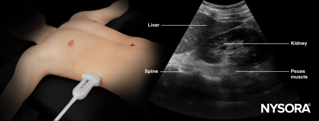
- Scan each kidney in longitudinal and transverse planes
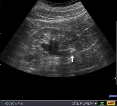
- Identify the renal cortex, medullary pyramids, and central collecting system
- Fan through the entire kidney
- Compare both sides
- Scan the bladder when obstruction is suspected
- Color Doppler can help distinguish vessels from a dilated collecting system
Hydronephrosis Severity
| Severity | Ultrasound Appearance |
|---|---|
| None | No dilation of the renal pelvis or calyces |
| Mild | Dilation of the renal pelvis with little or no calyceal dilation |
| Moderate | Dilation of the renal pelvis and calyces with preserved renal parenchyma |
| Severe | Marked dilation of the renal pelvis and calyces with cortical or parenchymal thinning |
Mild Hydronephrosis
- Renal pelvis dilated
- Calyces minimally dilated or normal
- Renal architecture preserved
- No cortical thinning
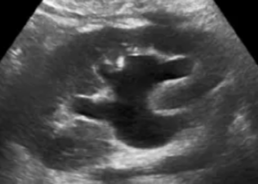
Moderate Hydronephrosis
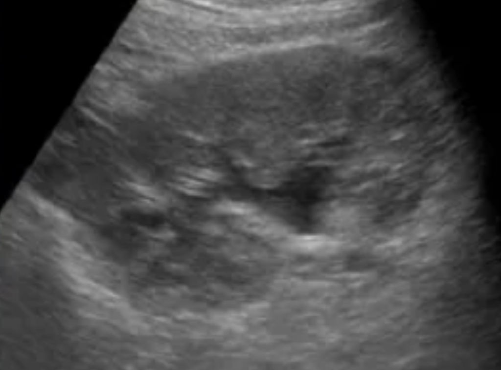
- Renal pelvis clearly dilated
- Calyces dilated
- Calyces become rounded or ballooned
- Renal parenchyma remains preserved
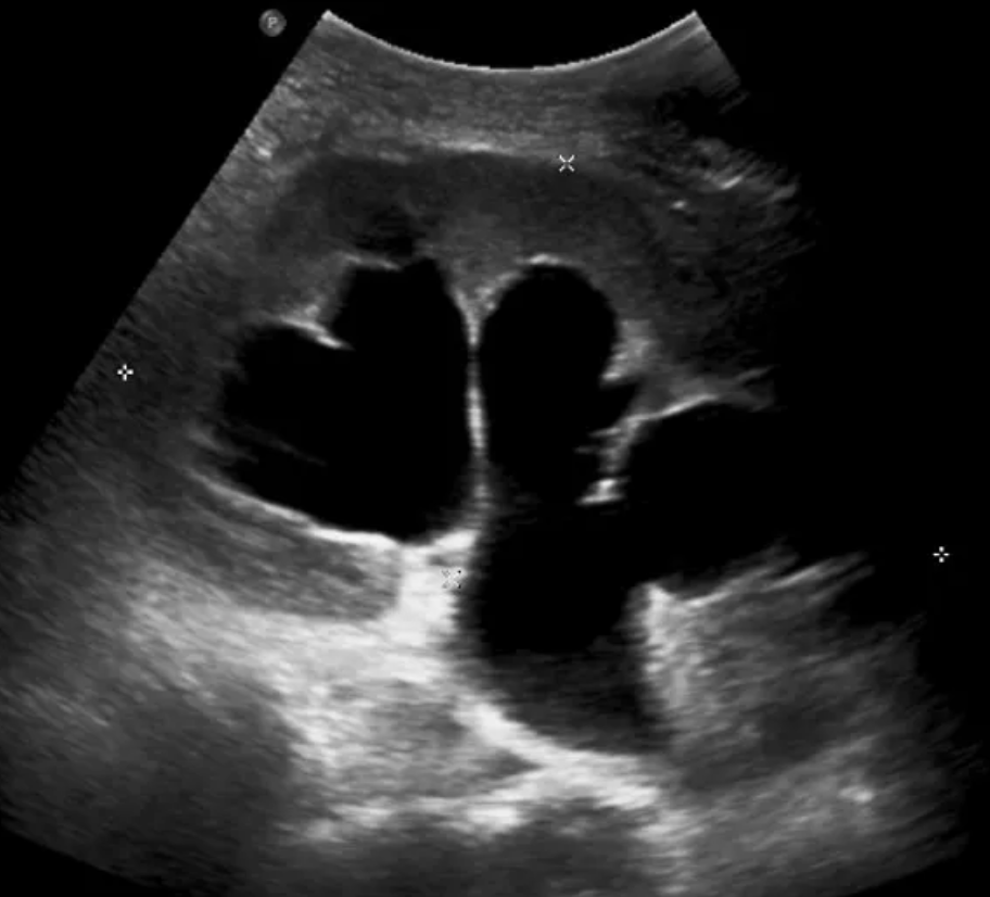
Severe Hydronephrosis
- Marked dilation of pelvis and calyces
- Ballooned calyces
- Loss of normal collecting system architecture
- Renal cortical or parenchymal thinning
Interpretation
| Ultrasound Pattern | Interpretation |
|---|---|
| Renal pelvic dilation with minimal or no calyceal dilation | Mild hydronephrosis |
| Renal pelvic and calyceal dilation with preserved parenchyma | Moderate hydronephrosis |
| Marked pelvic and calyceal dilation with parenchymal thinning | Severe hydronephrosis |
| Unilateral hydronephrosis with renal colic | Ureteral obstruction likely |
| Bilateral hydronephrosis | Consider distal obstruction |
| Hydronephrosis with a distended bladder | Consider bladder outlet obstruction |
| No hydronephrosis with suspected renal colic | Stone not excluded |
Full Bladder
A very full bladder or high fluid intake can transiently produce collecting-system dilation. Repeating the scan after voiding can be useful when the finding is equivocal.
Stones
- Hydronephrosis
- Echogenic stone if directly visualized
- Posterior acoustic shadowing
- Twinkle artifact on color Doppler
- Stone may be visible at the UPJ or UVJ