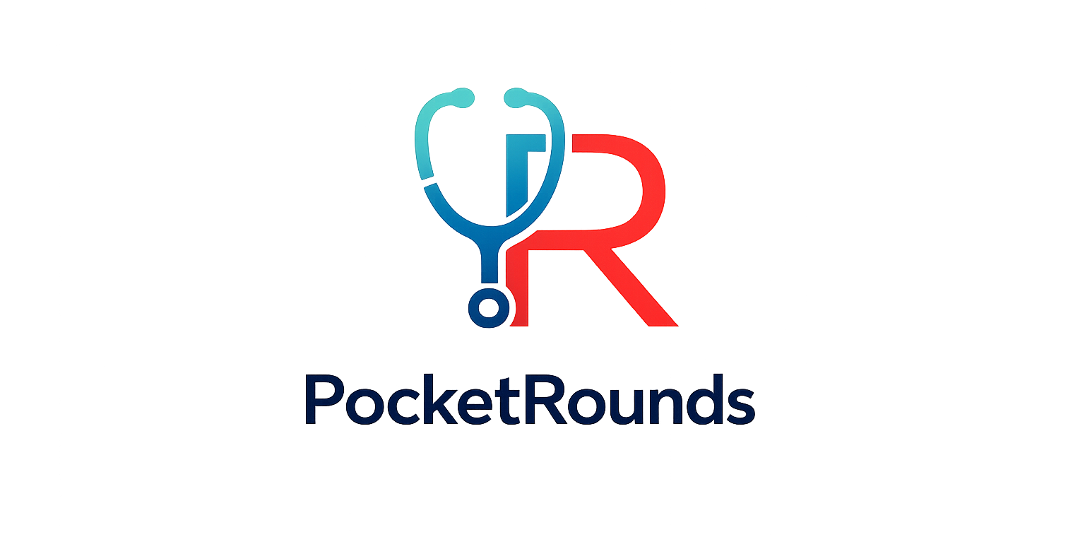
How to Scan
- Curvilinear abdominal probe
- Start subcostally in the RUQ
- Scan the entire gallbladder in longitudinal and transverse planes
- Follow the fundus, body, neck, and infundibulum
- Reposition into left lateral decubitus or upright position to assess mobility of stones or sludge
- Fasting for 6 to 8 hours is preferred when possible
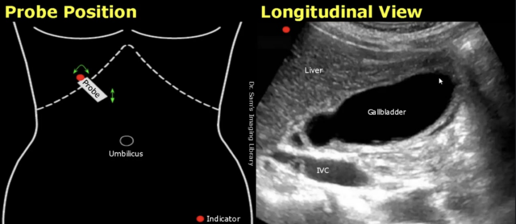
Normal Measurements
| Measurement | Normal | Abnormal |
|---|---|---|
| Wall thickness | < 3 mm | > 3 mm |
| Length | < 10 cm | ≥ 10 cm |
| Width | < 4 cm | ≥ 4 cm |
| Depth | < 4 cm | ≥ 4 cm |
Gallstones
- Echogenic focus
- Posterior acoustic shadowing
- Usually mobile with repositioning
- Ultrasound sensitivity >95% for gallstones
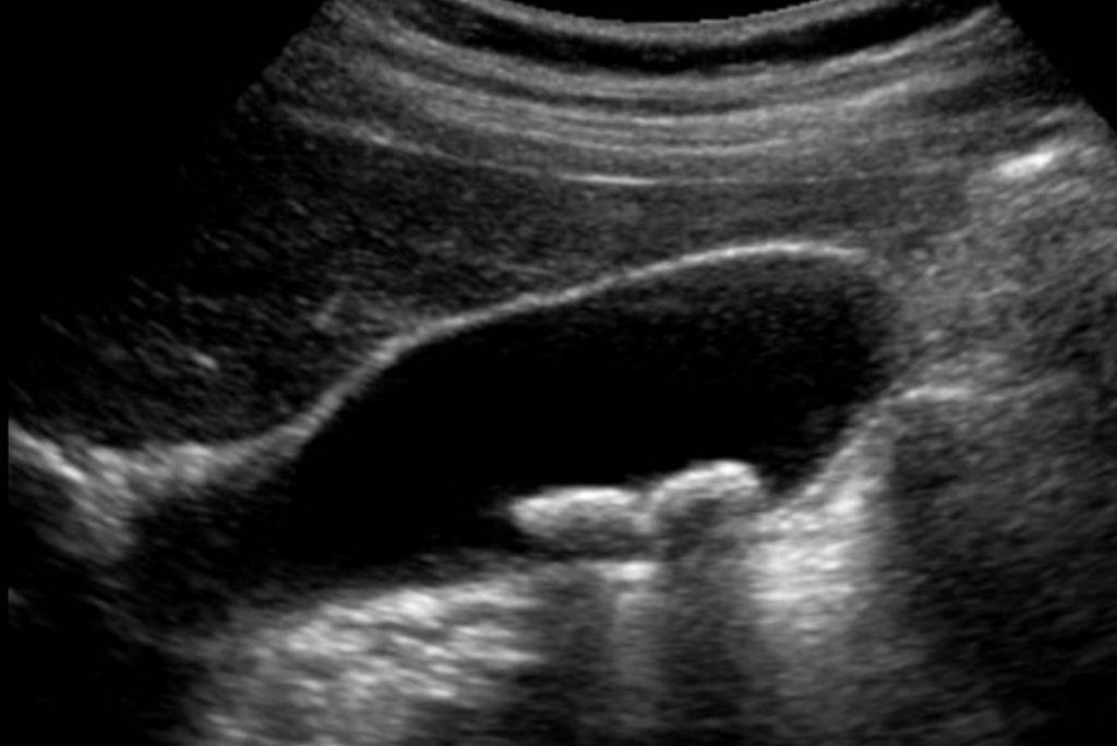
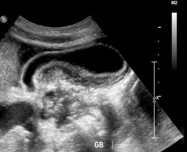
WES Sign
Wall Echo Shadow- Suggests GB packed with stones
- Gallbladder wall
- Echogenic stone surface
- Posterior acoustic shadow
Sludge
- Low-level internal echoes
- Usually dependent
- Moves slowly with repositioning
- No posterior acoustic shadowing
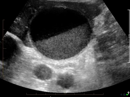
Gallbladder Polyp
- Fixed wall-attached lesion
- Usually does not shadow
- Does not move with repositioning
- Sludge typically moves while a true polyp remains fixed
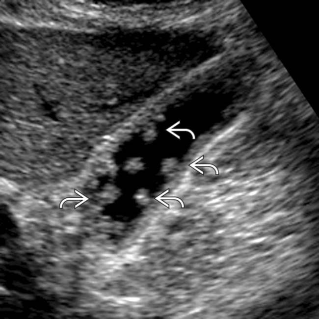
Adenomyomatosis
- Focal or diffuse wall thickening
- Intramural echogenic foci
- Comet-tail artifact
- Small intramural cystic spaces may be seen
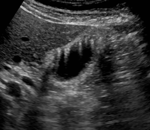
Stone vs. Sludge vs. Polyp vs. Adenomyomatosis?
| Finding | Appearance | Moves | Shadows |
|---|---|---|---|
| Gallstone | Echogenic focus | Usually | Yes |
| Sludge | Dependent low-level echoes | Usually slowly | No |
| Polyp | Wall-attached lesion | No | Usually no |
| Adenomyomatosis | Wall thickening with echogenic foci | No | Comet-tail artifact |
Acute Cholecystitis
Look for a combination of findings
- Gallstones
- Impacted stone at the neck or infundibulum
- Gallbladder wall >3 mm
- Gallbladder distention
- Sonographic Murphy sign
- Pericholecystic fluid
- Layered or edematous wall
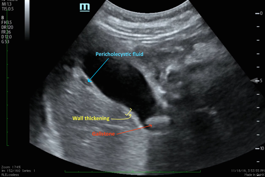
Wall Thickening Is Nonspecific
Can also occur with
- Right heart failure
- Cirrhosis
- Ascites
- Hypoalbuminemia
- Acute hepatitis
- Postprandial gallbladder contraction
Acalculous Cholecystitis
Consider especially in critically ill patients.
- Wall thickening
- Gallbladder distention
- Pericholecystic fluid
- Sludge or debris
- No gallstone required
Quick Interpretation
| Ultrasound Pattern | Likely Finding |
|---|---|
| Mobile shadowing stone with otherwise normal gallbladder | Cholelithiasis |
| Stone with wall thickening, distention, Murphy sign, or pericholecystic fluid | Acute calculous cholecystitis |
| Inflammatory gallbladder findings without stones in a critically ill patient | Acalculous cholecystitis |
| Dependent nonshadowing material that moves slowly | Biliary sludge |
| Fixed wall-attached nonshadowing lesion | Gallbladder polyp |
| Wall thickening with comet-tail artifact | Adenomyomatosis |
| Wall thickening without focal gallbladder inflammatory findings | Consider systemic cause |
References
Lucius C, Braden B, Jenssen C, et al.
Ultrasound of the Gallbladder: An Update on Measurements, Reference Values, Variants and Frequent Pathologies.
Life. 2025;15(6):941.
View article
American College of Radiology.
ACR Appropriateness Criteria® Right Upper Quadrant Pain.
American College of Radiology.
View guideline
Yokoe M, Hata J, Takada T, et al.
Tokyo Guidelines 2018: Diagnostic Criteria and Severity Grading of Acute Cholecystitis.
J Hepatobiliary Pancreat Sci. 2018;25(1):41-54.
View article
Pisano M, Allievi N, Gurusamy K, et al.
2020 World Society of Emergency Surgery Updated Guidelines for the Diagnosis and Treatment of Acute Calculous Cholecystitis.
World J Emerg Surg. 2020;15:61.
View guideline
Huang SS, Lin KW, Wu YT, et al.
Diagnostic Performance of Ultrasound in Acute Cholecystitis: A Systematic Review and Meta-analysis.
World J Emerg Surg. 2023;18:54.
View article