
Carotid Stenosis Severity
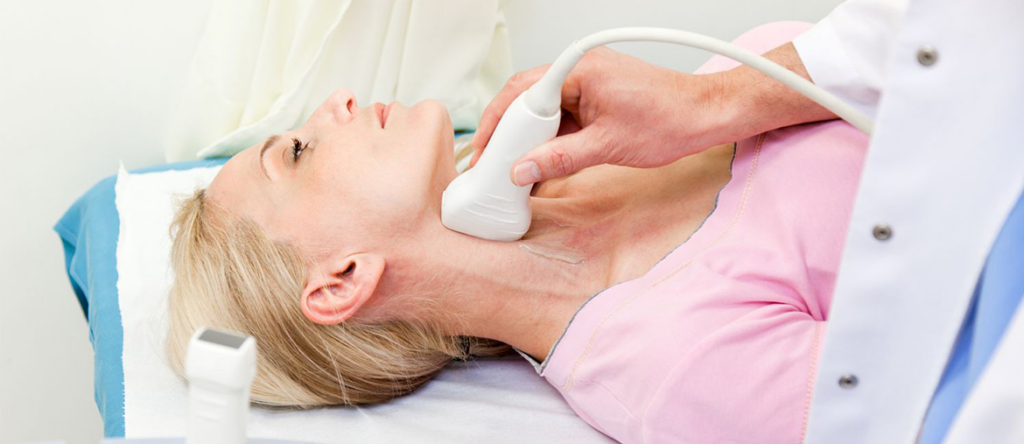
Setup
- High-frequency linear probe
- Patient supine, neck relaxed
- Head turned ~45 degrees away from side being scanned
- Scan CCA, bulb, ICA, ECA, and vertebral artery
- Use anterior, lateral, and posterolateral windows as needed
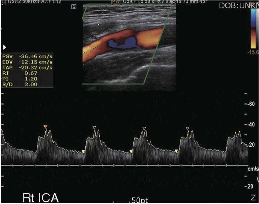
ICA
- Usually posterior/lateral
- Larger
- No branches
- Low-resistance waveform
- More diastolic flow
- No temporal tap artifact
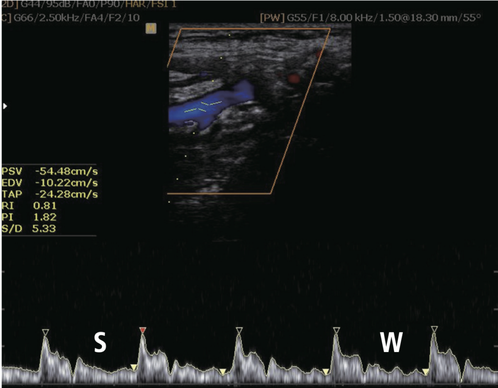
ECA
- Usually anterior/medial
- Smaller
- Has branches
- Higher-resistance waveform
- Less diastolic flow
- Temporal tapping causes waveform serrations
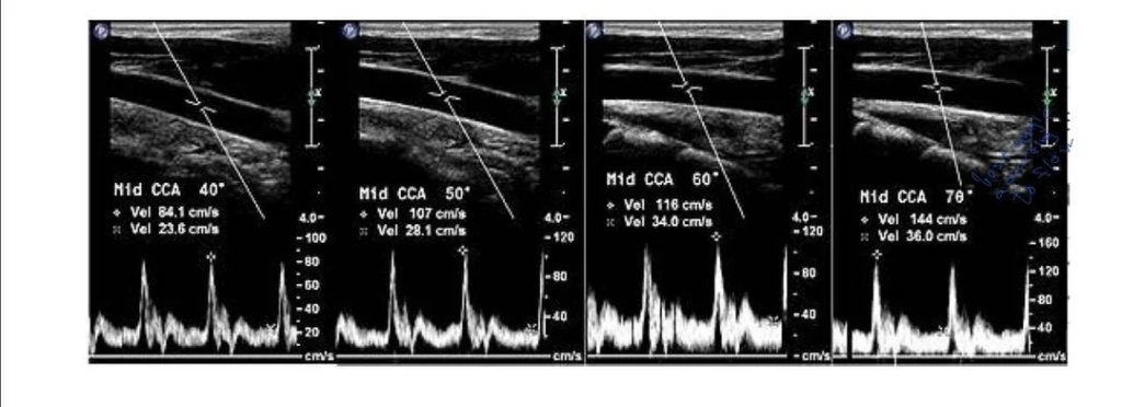
How to scan
Grayscale
- Evaluate CCA, bulb, ICA, and ECA in long and short axis
- Look for plaque, calcification, luminal narrowing, ulceration, and shadowing
- Describe plaque as echogenic, echolucent, heterogeneous, calcified, smooth, irregular, or ulcerated
Color Doppler
- Identify stenotic jet, aliasing, color bruit, residual lumen, or trickle flow
- Use color to find the tightest lesion, not to grade severity by itself
Spectral Doppler
- Keep angle correction ≤60 degrees
- Align cursor with flow direction
- Sample CCA and ICA
- Measure highest reliable ICA PSV at the stenotic jet
- Record ICA EDV and ICA/CCA PSV ratio
- Sample vertebral artery and document antegrade vs retrograde flow
Interpretation
- No plaque + normal velocities = normal
- Plaque + normal/mild velocity elevation = usually <50% stenosis
- ICA PSV is the main number, but do not use it alone
- Use EDV and ICA/CCA ratio when PSV is borderline or flow is altered
- Near occlusion can have high, low, or absent velocities
- Total occlusion requires no visible lumen and no flow on color, spectral, or power Doppler
ICA Stenosis Grading by Ultrasound
| ICA stenosis | ICA PSV | Plaque / grayscale estimate | ICA/CCA PSV ratio | ICA EDV |
|---|---|---|---|---|
| Normal | <180 cm/s | No plaque | <2.0 | <40 cm/s |
| <50% | <180 cm/s | Plaque present, <50% narrowing | <2.0 | <40 cm/s |
| 50-69% | 180-230 cm/s | >50% plaque/narrowing | 2.0-4.0 | 40-100 cm/s |
| >70% but less than near occlusion | >230 cm/s | >50% plaque/narrowing | >4.0 | >100 cm/s |
| Near occlusion | High, low, or undetectable | Marked luminal narrowing, trickle flow/string sign | Variable | Variable |
| Total occlusion | Undetectable | No detectable patent lumen | Not applicable | Not applicable |