ECMO Troubleshooter
Select the circuit and bedside problem. The interpretation updates automatically.
Bedside assessment
ECMO mode
Choose VV or VA support
Bedside problem
Problems change based on the ECMO mode
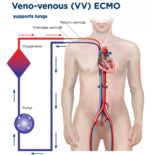
When to choose VV ECMO?
- Severe, potentially reversible respiratory failure (ARDS) refractory to optimal management
- Hypoxemia or hypercapnia despite lung-protective ventilation, proning, paralysis, or inhaled vasodilator
- Intact or manageable hemodynamics without need for circulatory support
- Typical setup: femoral–IJ or dual-lumen cannulation
- Targets: SaO₂ > 88–92%
- Goal: Rest the lung
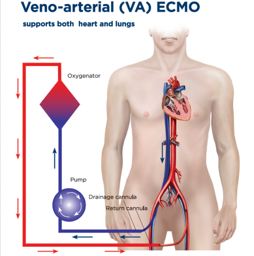
When to choose VA ECMO?
- Cardiogenic shock or cardiac arrest with potential for recovery or bridge
- Severe biventricular failure or refractory VT/VF, post-cardiotomy shock
- Need for hemodynamic support (MAP/perfusion) beyond vasoactives
- Typical setup: femoral VA
- Watch for: LV distension, Harlequin (north–south) syndrome
- Adjuncts: vent, Impella, or IABP if needed
Contraindications
- Irreversible disease without bridge or exit plan
- Multi-organ failure without recovery path
- Prohibitive bleeding/coagulopathy, devastating neuro injury
- Prolonged no-flow/low-flow arrest, advanced frailty, or poor baseline
Initial setup & targets
VV ECMO (oxygenation/CO₂)
- Blood flow ~5 L/min (up to 7–8). Aim > 60–70% of CO for SpO₂ > 90%
- Sweep 2–3 L/min, titrate to pCO₂
- Vent: tidal 3–4 mL/kg IBW, Pplat ≤ 25, PEEP individualized, FiO₂ minimal
- Goals: SpO₂ 88–92% (ok 85–88% if perfusion OK), pH > 7.25
Pearl: if sats low, check recirculation → flow → Hb/SaO₂/oxygenator
VA ECMO (perfusion/oxygenation)
- Flow titrated to MAP and end-organ perfusion. Treat LV distension early
- Monitor right radial ABG for Harlequin syndrome; add vent or V-A-V if needed
- Minimize catecholamines; optimize preload/afterload; unload LV if required
Anticoagulation
Heparin
- UFH unless bleeding risk → low-dose or no-anticoagulation strategy
- Anti-Xa preferred; aPTT/ACT adjunctive
- Anti-Xa: 0.3–0.7 IU/mL
- aPTT: 50–70 s
- ACT: 160–180 s
- Platelets: > 75–100 K
- Fibrinogen: > 150–200 mg/dL
Reassess after circuit change, bleeding, or oxygenator dysfunction.
ECMO Troubleshooting
| Problem | Likely causes | Key actions |
|---|---|---|
| Low SpO₂ VV ECMO |
|
|
| Hypercapnia |
|
|
| Hypotension |
|
|
| Drainage insufficiency |
|
|
| Oxygenator failure / high ΔP |
|
|
| Harlequin syndrome VA ECMO |
|
|
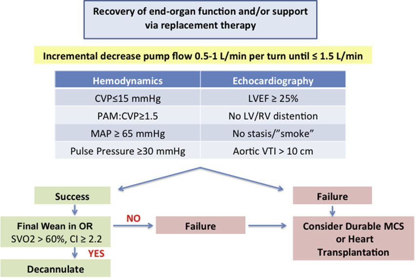
Weaning
VV ECMO
- Improving lung mechanics/oxygenation, minimal sweep
- Sweep-down or clamp test with permissive targets
VA ECMO
- Recovery of native CO: increasing pulse pressure, less inotrope, improving echo (LVOT VTI, aortic valve opening)
- Stepwise flow reduction under echo and hemodynamic monitoring
Daily checklist
- Indication still valid; exit strategy defined
- Flows, sweep, vent targets documented
- Anticoagulation and labs in range; hemolysis markers trended
- Limb/neuro checks, cannula inspection, circuit visual check
- Echo/ultrasound as needed for recirculation, function, effusions
References
- Combes, A., Peek, G. J., Hajage, D., Hardy, P., Abrams, D., Schmidt, M., Dechartres, A., Elbourne, D., & ECMO for Severe ARDS Systematic Review and Individual Patient Data Meta-analysis Investigators. (2020). ECMO for severe ARDS: Systematic review and individual patient data meta-analysis. Intensive Care Medicine, 46(11), 2048–2057. https://doi.org/10.1007/s00134-020-06248-3
- Combes, A., Hajage, D., Capellier, G., Demoule, A., Lavoué, S., Guervilly, C., Da Silva, D., Zafrani, L., Tirot, P., Veber, B., Maury, E., Levy, B., Cohen, Y., Richard, C., Kalfon, P., Bouadma, L., Mehdaoui, H., Beduneau, G., Lebreton, G., ... EOLIA Trial Group. (2018). Extracorporeal membrane oxygenation for severe acute respiratory distress syndrome. New England Journal of Medicine, 378(21), 1965–1975. https://doi.org/10.1056/NEJMoa1800385
- Peek, G. J., Mugford, M., Tiruvoipati, R., Wilson, A., Allen, E., Thalanany, M. M., Hibbert, C. L., Truesdale, A., Clemens, F., Cooper, N., Firmin, R. K., Elbourne, D., & CESAR Trial Collaboration. (2009). Efficacy and economic assessment of conventional ventilatory support versus extracorporeal membrane oxygenation for severe adult respiratory failure: A multicentre randomised controlled trial. The Lancet, 374(9698), 1351–1363. https://doi.org/10.1016/S0140-6736(09)61069-2
- Makdisi, G., & Wang, I. W. (2015). Extra corporeal membrane oxygenation review of a lifesaving technology. Journal of Thoracic Disease, 7(7), E166–E176. https://doi.org/10.3978/j.issn.2072-1439.2015.07.17
- Extracorporeal Life Support Organization. (2021). ELSO guidelines for adult respiratory failure. ASAIO Journal, 67(5), 465–495. https://doi.org/10.1097/MAT.0000000000001430
- Lorusso, R., Shekar, K., MacLaren, G., Schmidt, M., Pellegrino, V., Meyns, B., Haft, J., Vercaemst, L., Pappalardo, F., Mueller, T., Burrell, A., Babatasi, G., Rycus, P., Barbaro, R. P., Thiagarajan, R. R., & ELSO Interim Guideline Writing Group. (2021). ELSO interim guidelines for venoarterial extracorporeal membrane oxygenation in adult cardiac patients. ASAIO Journal, 67(8), 827–844. https://doi.org/10.1097/MAT.0000000000001510
- Extracorporeal Life Support Organization. (2021). ELSO anticoagulation guideline. Extracorporeal Life Support Organization. https://www.elso.org/ecmo-resources/elso-ecmo-guidelines.aspx