
Simplified IBD US Severity Assessment
Based on bowel wall thickness, doppler activity, wall stratification, and inflammatory fat
Generalized Severity Grading
Start
Suspected active IBDPerform ultrasound
↓
Step 1
BWT ≥3 mm?
↓
No
Inactive / remission
Yes
Proceed
↓
Step 2
Doppler ≥2?
↓
No
Mild disease
Yes
Proceed
↓
Step 3
Loss of stratificationand/or inflammatory fat?
↓
Partial
Moderate disease
Yes
Severe disease
↓
Override
Stricture, fistula, abscess,phlegmon, dilation?
↓
Yes
Severe / complicatedHow to do it
- Fasting ~6–8 hrs (optional but improves views)
- Supine positioning
- Use curvilinear (deep) + linear (high-res) probes
- Scan systematically: terminal ileum → proximally or full abdominal sweep
What you measure
- Bowel wall thickness (BWT)
- Doppler vascularity
- Wall stratification
- Peristalsis + compressibility
- Luminal narrowing / dilation
- Extramural findings (fat, nodes, abscess)
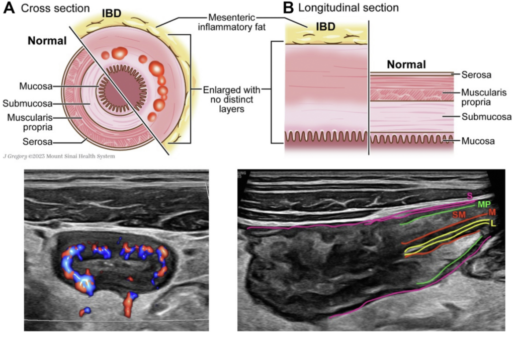
Bowel wall layers
- Mucosa → submucosa → muscularis → serosa
Bowel wall thickness
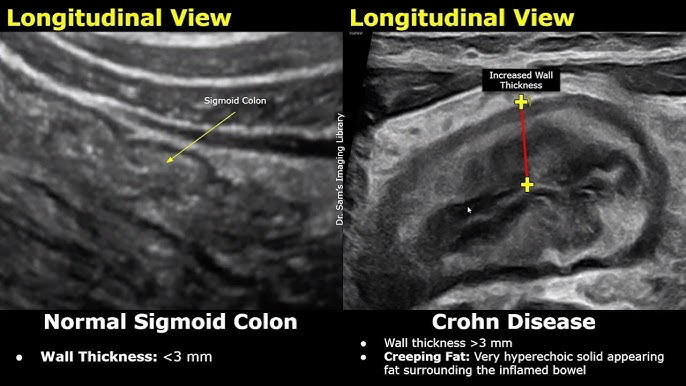
- Measure perpendicular to wall
- Longitudinal + transverse views
- Use most abnormal segment
- Normal: <3 mm
- Active inflammation: ≥3 mm
Doppler grading (Limberg)
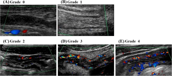
In general ≥2 = active inflammation
- 0 → no flow
- 1 → thickened wall, no flow
- 2 → spotty flow
- 3 → diffuse intramural flow
- 4 → flow into mesentery
Bowel wall stratification (BWS)
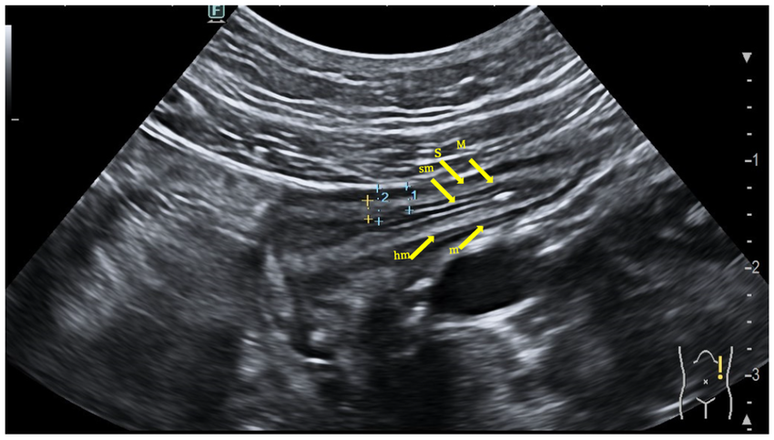
Loss of stratification = deeper, transmural inflammation
- 0 → normal (clear layered pattern)
- 1 → uncertain
- 2 → focal loss ≤3 cm
- 3 → extensive loss >3 cm
Inflammatory fat (i-fat)
Mesenteric fat hypertrophy adjacent to inflamed bowel (“creeping fat”). Appears hyperechoic surrounding affected segment. It is a marker of transmural inflammation and more common in Crohn's disease. Associated with stricturing and penetrating disease.
- 0 → absent
- 1 → uncertain
- 2 → present
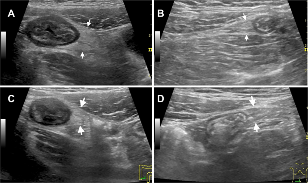
Features of active disease
- BWT ≥3 mm
- Increased Doppler signal
- Loss of wall stratification
- Inflammatory fat (creeping fat)
- Mesenteric lymph nodes
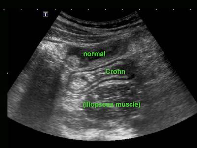
Validated Scoring systems
Ulcerative Colitis
- Milan Ultrasound Criteria (MUC)
- MUC = 1.4 × BWT + 2 × Doppler
- >6.2 → active disease
- UC-IUS index (0-7)
- BWT
- Doppler
- Haustration
- Fat
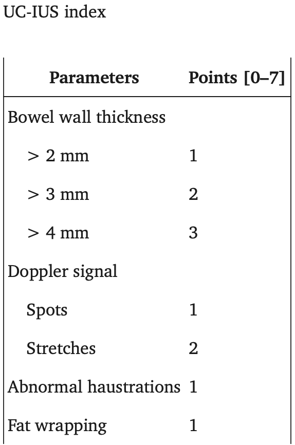
Crohn’s disease
- IBUS-SAS
- BWT
- BWS
- CDS
- I-fat
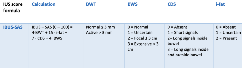
References
- Kucharzik, T., Tielbeek, J., Carter, D., Taylor, S. A., Tolan, D., Wilkens, R., Bryant, R. V., Maconi, G., Maaser, C., Panes, J., Rimola, J., Rieder, F., & Sturm, A. (2025). ECCO-ESGAR-ESP-IBUS guideline on diagnostics and monitoring of inflammatory bowel disease. Journal of Crohn’s and Colitis. https://doi.org/10.1093/ecco-jcc/jjaf108
- Maaser, C., Sturm, A., Vavricka, S. R., Kucharzik, T., Fiorino, G., Annese, V., Calabrese, E., Baumgart, D. C., Bettenworth, D., Borralho Nunes, P., Burisch, J., Castiglione, F., Eliakim, R., Ellul, P., González-Lama, Y., Gordon, H., Halligan, S., Katsanos, K., Kopylov, U., ... Stoker, J. (2019). ECCO-ESGAR guideline for diagnostic assessment in IBD part 1: Initial diagnosis, monitoring of known IBD, detection of complications. Journal of Crohn’s and Colitis, 13(2), 144–164. https://doi.org/10.1093/ecco-jcc/jjy113
- Novak, K. L., Nylund, K., Maaser, C., Petersen, F., Kucharzik, T., Lu, C., Allocca, M., Maconi, G., Panaccione, R., Ghosh, S., & Wilkens, R. (2021). Expert consensus on optimal acquisition and development of the International Bowel Ultrasound Segmental Activity Score [IBUS-SAS]: A reliability and inter-rater variability study on intestinal ultrasonography in Crohn’s disease. Journal of Crohn’s and Colitis, 15(4), 609–616. https://doi.org/10.1093/ecco-jcc/jjaa216
- Bots, S., Nylund, K., Löwenberg, M., & Gecse, K. B. (2021). Intestinal ultrasound to assess disease activity in ulcerative colitis: Development of a novel UC-ultrasound index. Journal of Crohn’s and Colitis, 15(8), 1264–1271. https://doi.org/10.1093/ecco-jcc/jjab002
- Allocca, M., Filippi, E., Costantino, A., Bonovas, S., Fiorino, G., Furfaro, F., Peyrin-Biroulet, L., & Danese, S. (2021). Milan ultrasound criteria are accurate in assessing disease activity in ulcerative colitis: External validation. United European Gastroenterology Journal, 9(4), 438–442. https://doi.org/10.1177/2050640620980203
- Maaser, C., Petersen, F., Helwig, U., Fischer, I., Roessler, A., Rath, S., Lang, D., Kucharzik, T., & German IBD Study Group. (2020). Intestinal ultrasound for monitoring therapeutic response in patients with ulcerative colitis: Results from the TRUST&UC study. Gut, 69(9), 1629–1636. https://doi.org/10.1136/gutjnl-2019-319451