Mitral Stenosis Severity Calculator
Severity
Mitral Stenosis Severity
| Severity | Mitral Valve Area | Mean Gradient |
|---|---|---|
| Mild | > 1.5 cm² | < 5 mmHg |
| Moderate | 1.0-1.5 cm² | 5-10 mmHg |
| Severe | < 1.0 cm² | > 10 mmHg |
2D assessment
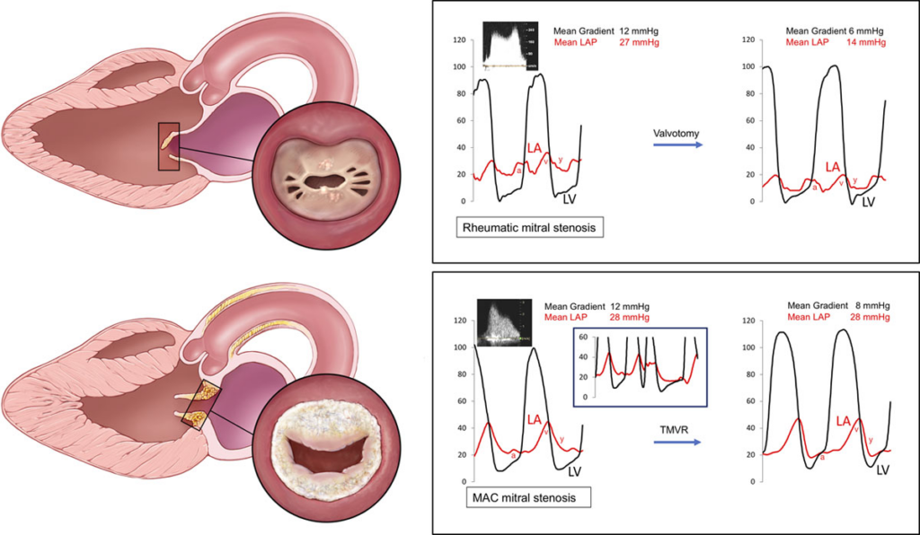
- Define etiology: rheumatic vs calcific
- Assess leaflet mobility, thickening, calcification, subvalvular disease
- Assess LA size, RV size/function, estimate RVSP
Planimetry
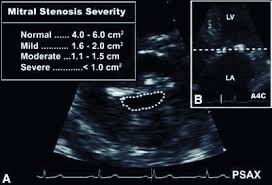
- Parasternal short-axis at leaflet tips
- Trace smallest orifice in mid-diastole
- Best direct measure of MVA if image quality adequate
Mean gradient
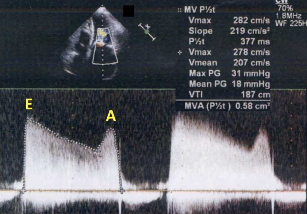
- CW Doppler from apical 4-chamber
- Trace full diastolic envelope
- Document heart rate and rhythm
- Gradient increases with tachycardia and high cardiac output
- This tracing will provide the mean gradient and MVA by PHT using continuity equation
- MVA = (LVOT area × LVOT VTI) / MV VTI
- To use if planimetry is not feasible