
Vascular Territory Prediction Model
Select EKG changes and echo wall abnormalities. The calculator estimates the most likely vascular territory and culprit artery, and suggests echo views to verify.
EKG findings
Check what you see.
Echo wall-motion findings
Regional hypokinesis or akinesis by wall.
Context
Reset clears all inputs.
Logic
Territory likelihood
Top territory reflects combined EKG and echo signals.
Likely culprit artery
Dominance and wrap-around variants can shift probability.
Why this result?
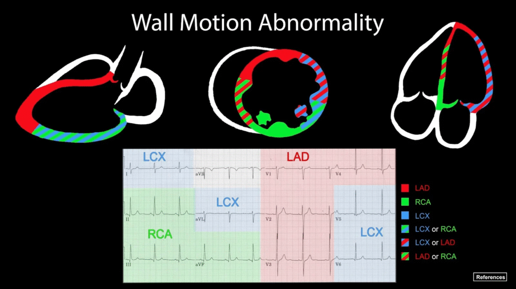
EKG → Coronary Artery → Echo Wall-Motion Segments
| EKG leads | Coronary Artery | Territory | TTE Wall Motion |
|---|---|---|---|
| V1–V2 (septal) | LAD (septal perforators; proximal LAD if large) | Anteroseptal | PSAX (pap level) for septum, A4C for septal wall, A3C for anteroseptal, APLAX for basal anteroseptal |
| V3–V4 (anterior) | LAD | Anterior/apex | A2C for anterior/apex, PSAX for anterior, A3C for anterior/apex |
| I, aVL (high lateral) | LCx (OM) or LAD (diagonal) | High lateral | PSAX for lateral wall, A3C for anterolateral, A4C for lateral mid/apex |
| V5–V6 (lateral) | LCx (OM) > LAD (diagonal) | Lateral | A4C for lateral wall/apex, PSAX for basal/mid lateral |
| II, III, aVF (inferior) | RCA (dominant) > LCx (if left dominant) | Inferior | A2C for inferior/apex, PSAX for basal/mid inferior |
| “Posterior” (ST↓ V1–V3; tall R V1–V2; STE V7–V9) | RCA (dominant) or LCx | Posterior/inferolateral | PSAX for posterior, A3C for inferolateral |
| V1–V4 with right-sided V4R STE | Proximal RCA | RV infarct (± inferior LV) | A4C/RV-focused views; TAPSE/TDI impaired |
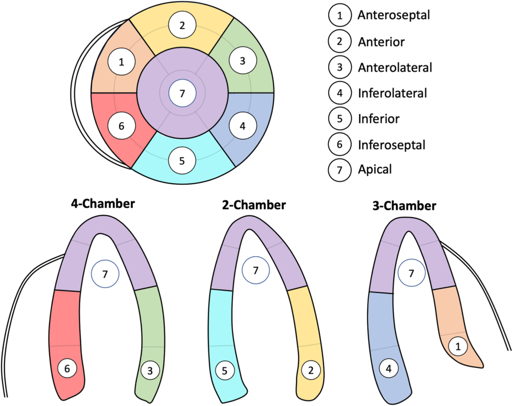
Echo view → wall cheat-sheet
| Echo view | Walls/segments primarily seen | Typical culprit when abnormal |
|---|---|---|
| A4C | Septal and lateral walls; apex | LAD (septal/anterior), LCx/Diagonal (lateral) |
| A2C | Inferior and anterior walls; apex | RCA/LCx (inferior), LAD (anterior) |
| A3C (Apical long-axis) | Anteroseptal and inferolateral walls; apex | LAD (anteroseptal), LCx/RCA (inferolateral) |
| PLAX | Basal/mid anteroseptal and inferolateral/posterior slices | LAD (anteroseptal), LCx/RCA (posterolateral) |
| PSAX (pap level) | Full circumferential slice of LV | LAD (anterior/septal), LCx/RCA (inferolateral/inferior) |
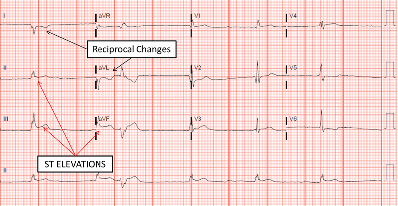
Reciprocal patterns
| STE territory | Common reciprocal ST depression |
|---|---|
| Anterior | Inferior leads |
| Lateral | Inferior leads |
| Inferior | High lateral leads (I, aVL) |
| Posterior | Anterior leads (appears as ST depression V1–V3) |
References
- Lang, R. M., Badano, L. P., Mor-Avi, V., Afilalo, J., Armstrong, A., Ernande, L., Flachskampf, F. A., Foster, E., Goldstein, S. A., Kuznetsova, T., Lancellotti, P., Muraru, D., Picard, M. H., Rietzschel, E. R., Rudski, L., Spencer, K. T., Tsang, W., & Voigt, J.-U. (2015). Recommendations for cardiac chamber quantification by echocardiography in adults: An update from the American Society of Echocardiography and the European Association of Cardiovascular Imaging. Journal of the American Society of Echocardiography, 28(1), 1–39.e14. https://doi.org/10.1016/j.echo.2014.10.003
- Cerqueira, M. D., Weissman, N. J., Dilsizian, V., Jacobs, A. K., Kaul, S., Laskey, W. K., Pennell, D. J., Rumberger, J. A., Ryan, T., & Verani, M. S. (2002). Standardized myocardial segmentation and nomenclature for tomographic imaging of the heart: A statement for healthcare professionals from the Cardiac Imaging Committee of the Council on Clinical Cardiology of the American Heart Association. Circulation, 105(4), 539–542. https://doi.org/10.1161/hc0402.102975
- Thygesen, K., Alpert, J. S., Jaffe, A. S., Chaitman, B. R., Bax, J. J., Morrow, D. A., & White, H. D. (2018). Fourth universal definition of myocardial infarction. Circulation, 138(20), e618–e651. https://doi.org/10.1161/CIR.0000000000000617
- Ibanez, B., James, S., Agewall, S., Antunes, M. J., Bucciarelli-Ducci, C., Bueno, H., Caforio, A. L. P., Crea, F., Goudevenos, J. A., Halvorsen, S., Hindricks, G., Kastrati, A., Lenzen, M. J., Prescott, E., Roffi, M., Valgimigli, M., Varenhorst, C., Vranckx, P., & Widimský, P. (2018). 2017 ESC guidelines for the management of acute myocardial infarction in patients presenting with ST-segment elevation. European Heart Journal, 39(2), 119–177. https://doi.org/10.1093/eurheartj/ehx393
- O’Gara, P. T., Kushner, F. G., Ascheim, D. D., Casey, D. E., Jr., Chung, M. K., de Lemos, J. A., Ettinger, S. M., Fang, J. C., Fesmire, F. M., Franklin, B. A., Granger, C. B., Krumholz, H. M., Linderbaum, J. A., Morrow, D. A., Newby, L. K., Ornato, J. P., Ou, N., Radford, M. J., Tamis-Holland, J. E., ... Zhao, D. X. (2013). 2013 ACCF/AHA guideline for the management of ST-elevation myocardial infarction. Circulation, 127(4), e362–e425. https://doi.org/10.1161/CIR.0b013e3182742cf6
- Birnbaum, Y., Drew, B. J., & The International Society for Computerized Electrocardiology. (2003). The electrocardiogram in ST elevation acute myocardial infarction: Correlation with coronary anatomy and prognosis. Postgraduate Medical Journal, 79(935), 490–504. https://doi.org/10.1136/pmj.79.935.490