
AS Severity Grading
| Parameter | Mild | Moderate | Severe |
|---|---|---|---|
| Mean gradient (mmHg) | <20 | 20–39 | ≥40 |
| AVA (cm²) | >1.5 | 1.0–1.5 | ≤1.0 |
| Dimensionless index (LVOT VTI / AV VTI) | >0.50 | 0.25–0.50 | <0.25 |
AS Calculator
Severe AVA ≤1.0 cm² or DI <0.25 | Low flow SVI <35 mL/m² | Low gradient <40 mmHg | Reduced EF <50%
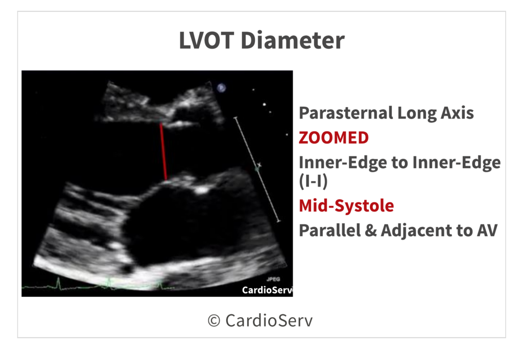
1. LVOT Diameter (PLAX)
- PLAX view
- Zoom on the aortic valve and LVOT
- Timing: Mid-systole (valve open)
- Measure inner-edge to inner-edge
- LVOT Area = 0.785 × (LVOT diameter)²
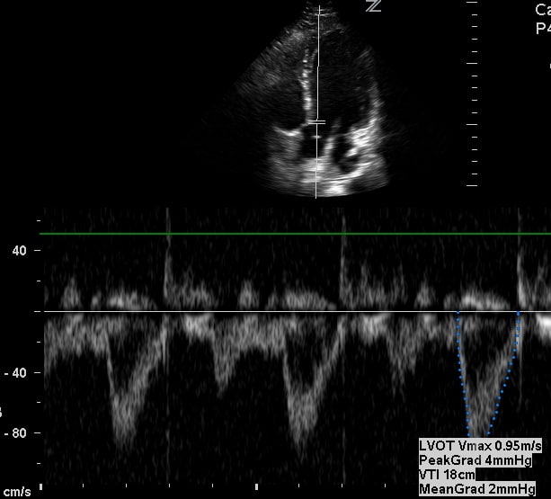
2. LVOT VTI (PW Doppler)
Represents forward stroke flow before the stenotic valve.
- Apical 5-chamber or 3-chamber
- PW doppler
- 0.5–1.0 cm below the aortic valve
3. Aortic Valve VTI (CW Doppler)
Measures velocity through the stenotic orifice
- CW Doppler
- Use multiple windows:
- Apical 5-chamber
- Apical 3-chamber
- Right parasternal
- Suprasternal notch
- Use the window with the highest reproducible velocity
- Trace the full systolic envelope
- Peak velocity (Vmax)
- Mean gradient
- AV VTI
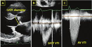
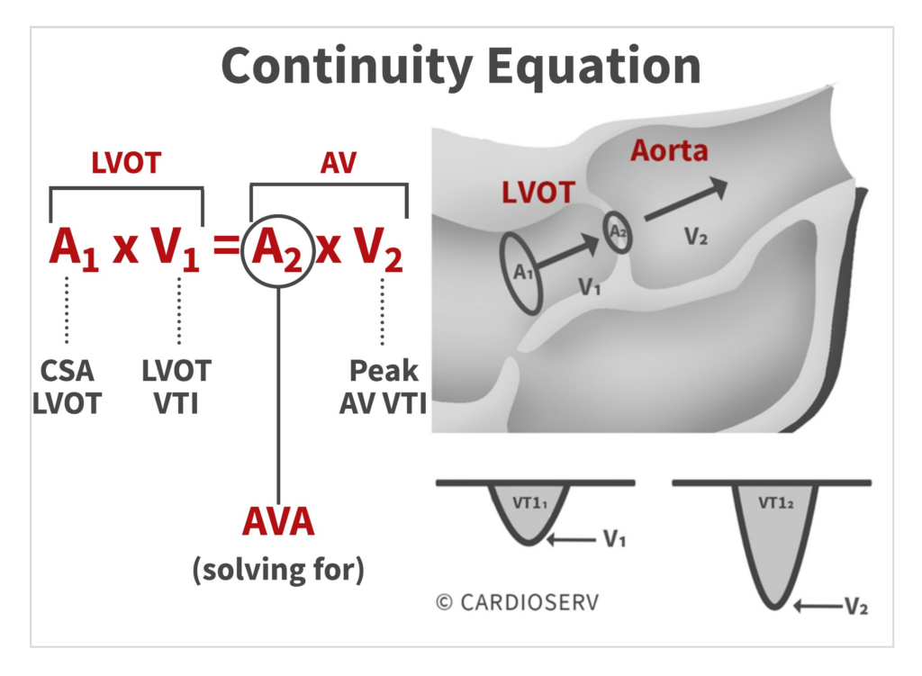
4. Aortic Valve Area (Continuity Equation)
AVA = (LVOT Area × LVOT VTI) ÷ AV VTI
5. Dimensionless Index (DI)
DI = LVOT VTI ÷ AV VTI
- ≤ 0.25 = Severe
- 0.25–0.50 = Moderate
- 0.50 = Mild
6. Gradients
Echo uses the Bernoulli equation:
- ΔP = 4V²
- Peak gradient = highest instantaneous pressure difference
- Mean gradient = average pressure difference across systole
7. Flow State
Stroke Volume = LVOT Area × LVOT VTI
- Index to BSA → SVI
- Low flow if:
- SVI < 35 mL/m²
- Low flow can cause low gradients even if valve is truly severe
References
- Baumgartner, H., Hung, J., Bermejo, J., Chambers, J. B., Evangelista, A., Griffin, B. P., Iung, B., Otto, C. M., Pellikka, P. A., & Quiñones, M. (2017). Recommendations on the echocardiographic assessment of aortic valve stenosis: A focused update from the European Association of Cardiovascular Imaging and the American Society of Echocardiography. Journal of the American Society of Echocardiography, 30(4), 372–392. https://doi.org/10.1016/j.echo.2017.02.009
- Otto, C. M., Nishimura, R. A., Bonow, R. O., Carabello, B. A., Erwin, J. P., III, Gentile, F., Jneid, H., Krieger, E. V., Mack, M., McLeod, C., O’Gara, P. T., Rigolin, V. H., Sundt, T. M., III, & Thompson, A. (2021). 2020 ACC/AHA guideline for the management of patients with valvular heart disease. Circulation, 143(5), e72–e227. https://doi.org/10.1161/CIR.0000000000000923
- Vahanian, A., Beyersdorf, F., Praz, F., Milojevic, M., Baldus, S., Bauersachs, J., Capodanno, D., Conradi, L., De Bonis, M., De Paulis, R., Delgado, V., Freemantle, N., Gilard, M., Haugaa, K. H., Jeppsson, A., Jüni, P., Pierard, L., Prendergast, B. D., Sádaba, J. R., ... Wojakowski, W. (2022). 2021 ESC/EACTS guidelines for the management of valvular heart disease. European Heart Journal, 43(7), 561–632. https://doi.org/10.1093/eurheartj/ehab395
- Nishimura, R. A., Otto, C. M., Bonow, R. O., Carabello, B. A., Erwin, J. P., III, Fleisher, L. A., Jneid, H., Mack, M. J., McLeod, C. J., O’Gara, P. T., Rigolin, V. H., Sundt, T. M., III, & Thompson, A. (2017). 2017 AHA/ACC focused update of the 2014 AHA/ACC guideline for the management of patients with valvular heart disease. Circulation, 135(25), e1159–e1195. https://doi.org/10.1161/CIR.0000000000000503
- Clavel, M. A., Magne, J., & Pibarot, P. (2016). Low-gradient aortic stenosis. European Heart Journal, 37(34), 2645–2657. https://doi.org/10.1093/eurheartj/ehw096
- Clavel, M. A., Messika-Zeitoun, D., Pibarot, P., Aggarwal, S. R., Malouf, J., Araoz, P. A., Michelena, H. I., Cueff, C., Larose, É., Capoulade, R., Vahanian, A., Enriquez-Sarano, M., & Dumesnil, J. G. (2013). The complex nature of discordant severe calcified aortic valve disease grading: New insights from combined Doppler echocardiographic and computed tomographic study. Journal of the American College of Cardiology, 62(24), 2329–2338. https://doi.org/10.1016/j.jacc.2013.08.1621