
Fascicular Block Algorithm
LAFB pattern
- LAD
- qR in I / aVL
- rS in II / III / aVF
- QRS normal or slightly wide
LPFB pattern
- RAD
- rS in I / aVL
- qR in II / III / aVF
- QRS usually normal
Is RBBB also present?
QRS ≥120 ms, rsR′ in V1, broad S in I / V6
Bifascicular block
RBBB + LAFB = most common
RBBB + LPFB = less common
RBBB + LPFB = less common
Isolated fascicular block
Isolated LAFB or isolated LPFB
PR >200 ms?
Trifascicular pattern
Bifascicular block + 1° AV block
Bifascicular block only
Pocket Guide
| Type | EKG Features | Notes |
|---|---|---|
| LAFB | LAD, qR in aVL, rS in II/III/aVF | Common, often benign |
| LPFB | RAD, rS in I/aVL, qR in II/III/aVF | Rare, usually structural disease |
| Bifascicular (RBBB + LAFB) | RBBB + LAD pattern | Most common bifascicular combo |
| Bifascicular (RBBB + LPFB) | RBBB + RAD pattern | Less common, consider ischemia |
| Trifascicular | Bifascicular + 1° AV block (PR >200 ms) | May progress to complete block |
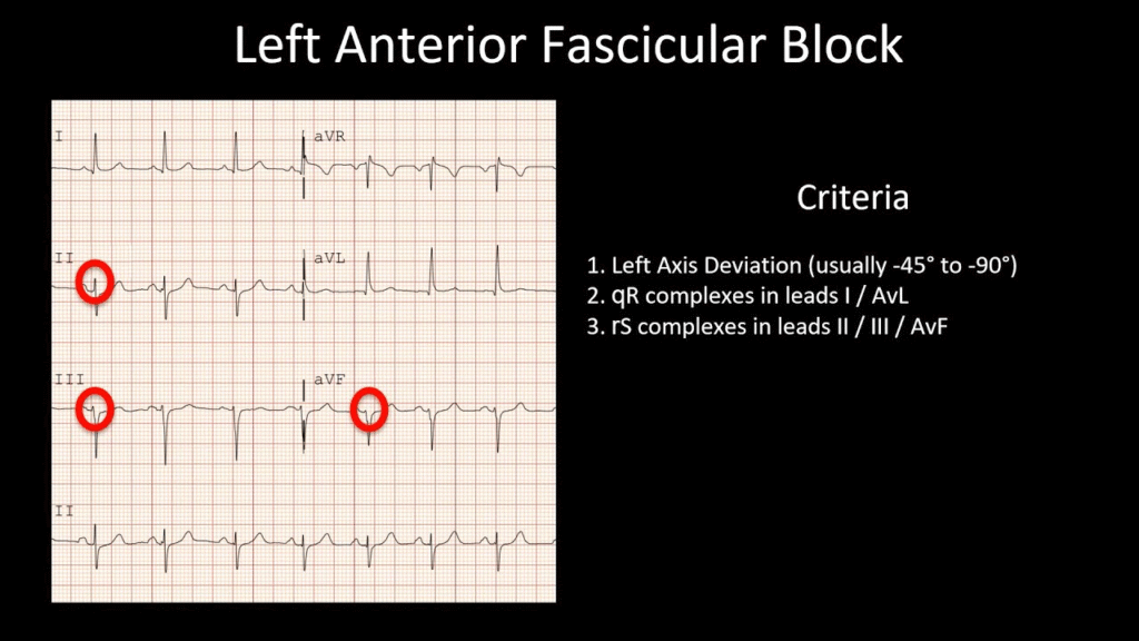
Left Anterior Fascicular Block (LAFB)
- Most common fascicular block
- EKG criteria:
- Left axis deviation (–45° to –90°)
- Small Q in leads I and aVL
- Small R in II, III, aVF
- Normal or slightly widened QRS (≤120ms)
- Often benign but may indicate underlying structural disease
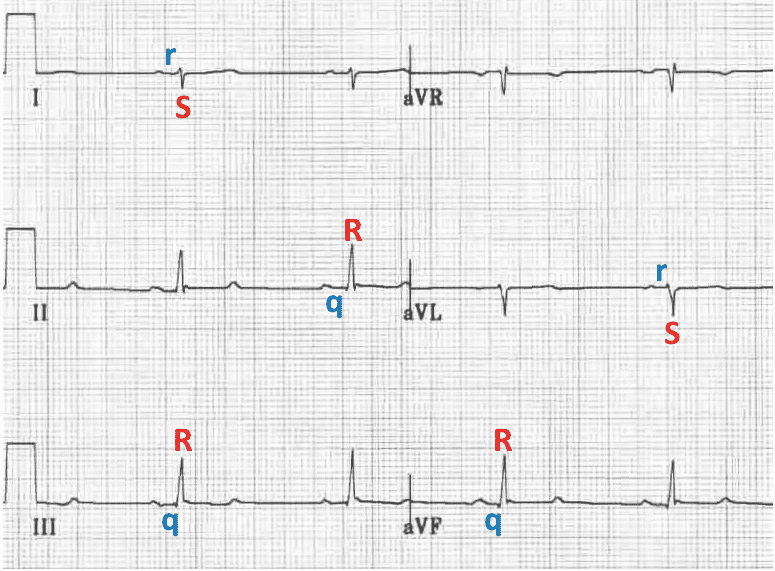
Left Posterior Fascicular Block (LPFB)
- Rare (posterior fascicle is broader, dual supply)
- EKG criteria:
- Right axis deviation (>+90°) without other cause (RVH, PE)
- Small R in I and aVL, small Q in II, III, aVF
- Normal QRS duration
- Suspect structural heart disease
Bifascicular Block
- Involves any two of the three fascicles:
- RBBB + LAFB (most common)
- RBBB + LPFB
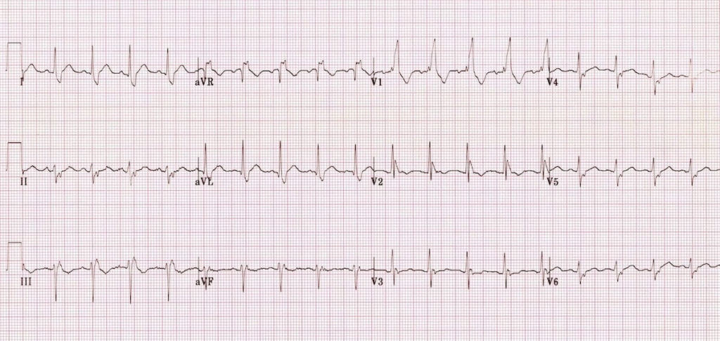
EKG
- RBBB: QRS ≥120 ms, rsR′ in V1, broad S in I/V6
- LAFB or LPFB criteria as above (in EKG below there is RBBB, LAD, normal QRS, rS complexes in inferior leads = RBBB + LAFB)
Clinical Significance
- Marker of conduction system disease
- Increases risk of progression to complete heart block, especially if:
- Alternating bundle branch block is present
- There's associated syncope
- May need pacemaker if symptomatic
Trifascicular Block
- Imprecise term; often refers to:
- RBBB + LAFB/LPFB + prolonged PR interval
- Not necessarily all 3 fascicles blocked simultaneously
- Consider EP study or pacing if symptomatic
References
- Surawicz, B., Childers, R., Deal, B. J., Gettes, L. S., Bailey, J. J., Gorgels, A., Hancock, E. W., Josephson, M., Kligfield, P., Kors, J. A., Macfarlane, P., Mason, J. W., Mirvis, D. M., Okin, P., Pahlm, O., Rautaharju, P. M., & van Herpen, G. (2009). AHA/ACCF/HRS recommendations for the standardization and interpretation of the electrocardiogram: Part III: Intraventricular conduction disturbances. Journal of the American College of Cardiology, 53(11), 976–981. https://doi.org/10.1016/j.jacc.2008.12.013
- Kusumoto, F. M., Schoenfeld, M. H., Barrett, C., Edgerton, J. R., Ellenbogen, K. A., Gold, M. R., Goldschlager, N. F., Hamilton, R. M., Joglar, J. A., Kim, R. J., Lee, R., Marine, J. E., McLeod, C. J., Oken, K. R., Patton, K. K., Pellegrini, C. N., Selzman, K. A., Thompson, A., & Varosy, P. D. (2019). 2018 ACC/AHA/HRS guideline on the evaluation and management of patients with bradycardia and cardiac conduction delay. Journal of the American College of Cardiology, 74(7), e51–e156. https://doi.org/10.1016/j.jacc.2018.10.044
- Pérez-Riera, A. R., Barbosa-Barros, R., Daminello-Raimundo, R., de Abreu, L. C., Baranchuk, A., & Nikus, K. (2018). Left posterior fascicular block, state-of-the-art review: A 2018 update. Indian Pacing and Electrophysiology Journal, 18(6), 217–230. https://doi.org/10.1016/j.ipej.2018.10.001
- Milliken, J. A., Macfarlane, P. W., & Lawrie, T. D. V. (1983). Isolated and complicated left anterior fascicular block: A review of suggested electrocardiographic criteria. Journal of Electrocardiology, 16(2), 121–128. https://doi.org/10.1016/S0022-0736(83)80018-1
- Elizari, M. V., Acunzo, R. S., & Ferreiro, M. (2007). Hemiblocks revisited. Circulation, 115(9), 1154–1163. https://doi.org/10.1161/CIRCULATIONAHA.106.637389