LVOT VTI Cardiac Output
Calculate stroke volume and cardiac output from LVOT diameter and pulsed-wave Doppler VTI.
LVOT Measurements
LVOT diameter
Inner-edge to inner-edge in mid-systole, typically from the parasternal long-axis view
cm
LVOT VTI
Trace the pulsed-wave Doppler envelope just proximal to the aortic valve
cm
Heart rate
Use the heart rate corresponding to the measured VTI
bpm
Cardiac Index
Calculate cardiac index
Adds body surface area and indexes cardiac output to body size
Height
cm
Weight
kg
Reference & Equations
| Parameter | Typical reference |
|---|---|
| LVOT VTI | Approximately 18–22 cm |
| Low LVOT VTI | <18 cm suggests reduced forward stroke distance |
| Stroke volume | Approximately 60–100 mL |
| Cardiac output | Approximately 4–8 L/min |
| Cardiac index | Approximately 2.5–4.0 L/min/m² |
| Low cardiac index | <2.2 L/min/m² is commonly used as a low-output threshold |
LVOT area = π × (LVOT diameter ÷ 2)²
Stroke volume = LVOT area × LVOT VTI
Cardiac output = stroke volume × heart rate ÷ 1000
BSA = √[(height × weight) ÷ 3600]
Cardiac index = cardiac output ÷ BSA
Stroke volume = LVOT area × LVOT VTI
Cardiac output = stroke volume × heart rate ÷ 1000
BSA = √[(height × weight) ÷ 3600]
Cardiac index = cardiac output ÷ BSA
LVOT VTI to Measure Cardiac Output
LVOT Area = π × (LVOT diameter / 2)²
Units
- Diameter in cm
- VTI in cm
- SV in mL
- CO in L/min
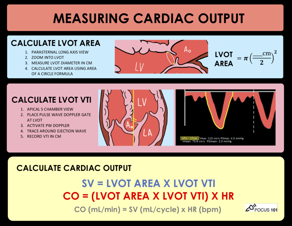
Step 1: Measure LVOT Diameter
View: Parasternal long axis
Timing: Mid systole
Method: Inner edge to inner edge
Location: Just proximal to the aortic valve annulus
Diameter is squared in the equation. Small measurement errors create HUGE CO errors.
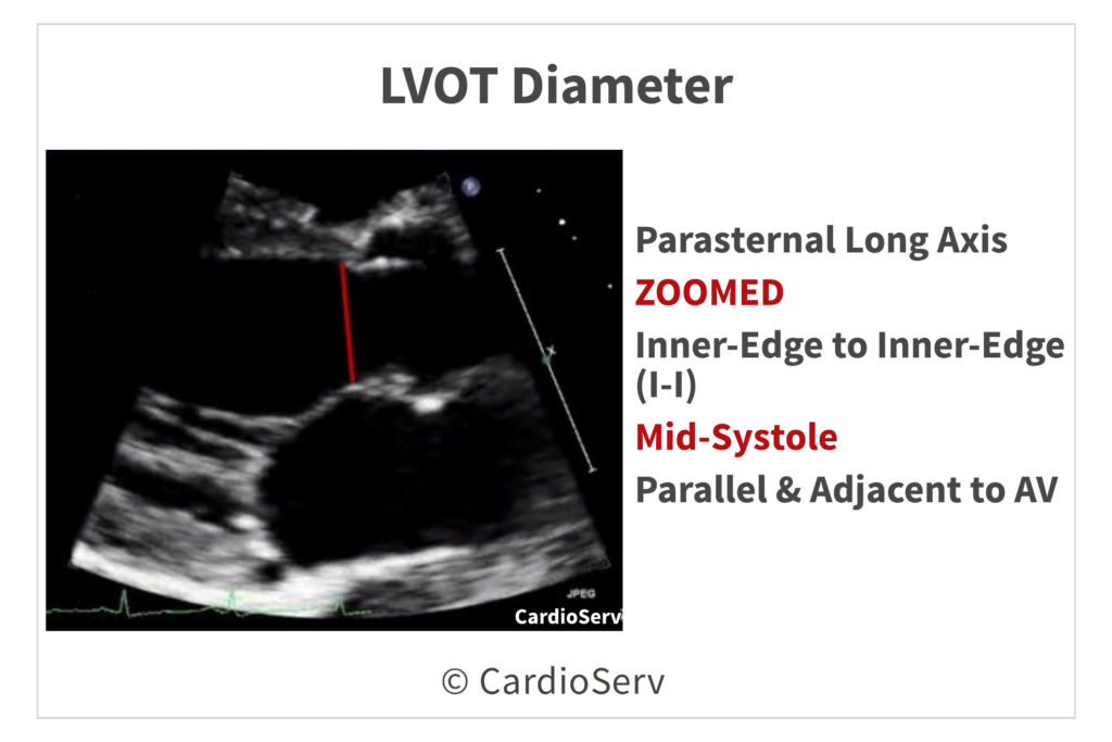
Step 2: Measure LVOT VTI
View: Apical 5 chamber (usually best) or apical 3 chamber
Doppler: Pulsed wave
How to do it
- Place PW Doppler sample gate 0.5 to 1.0 cm proximal to the aortic valve, on the LV side.
- Align Doppler beam as parallel to LVOT flow as possible.
- Increase sweep speed.
- Trace the outer edge of the Doppler envelope.
- Average:
- 3 beats in sinus rhythm
- 5 to 10 beats in atrial fibrillation
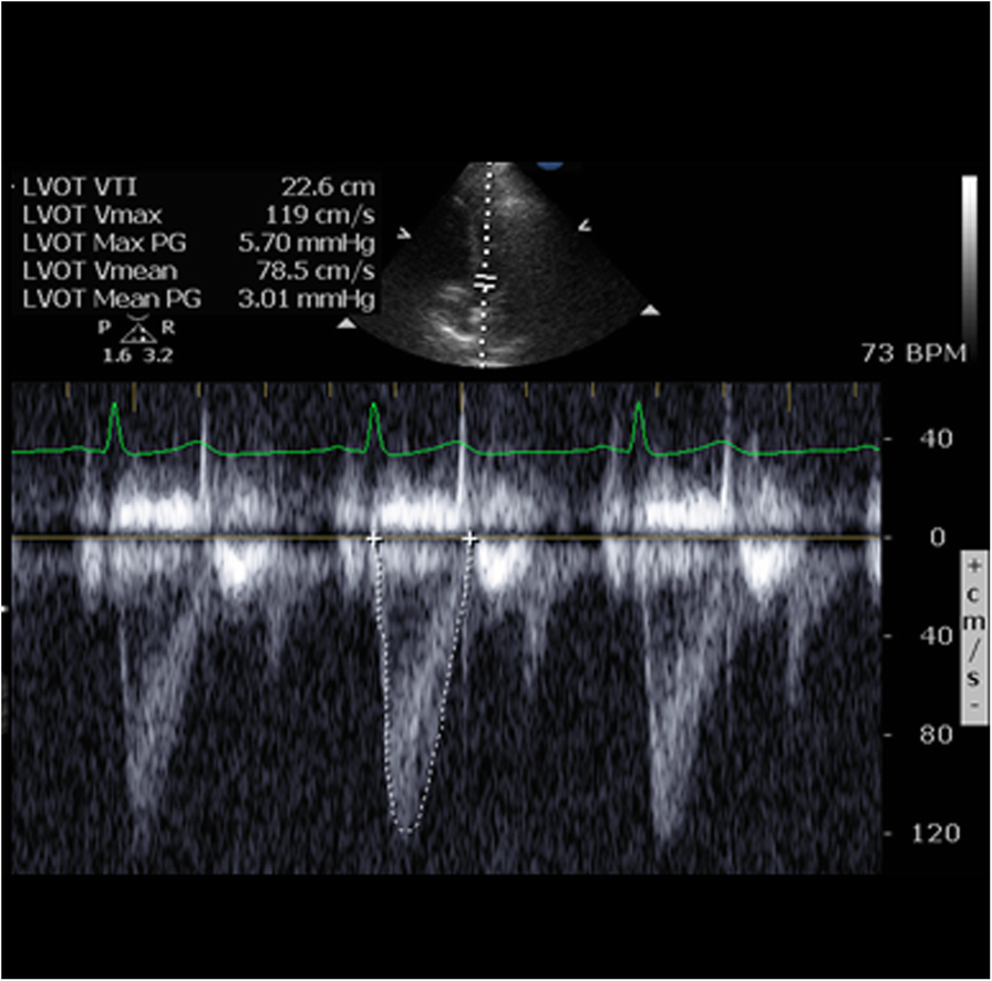
Step 3: Calculate Stroke Volume
- Stroke Volume = LVOT Area × LVOT VTI
Step 4: Calculate Cardiac Output
- CO = SV × HR
References
- Lang, R. M., Badano, L. P., Mor-Avi, V., Afilalo, J., Armstrong, A., Ernande, L., Flachskampf, F. A., Foster, E., Goldstein, S. A., Kuznetsova, T., Lancellotti, P., Muraru, D., Picard, M. H., Rietzschel, E. R., Rudski, L., Spencer, K. T., Tsang, W., & Voigt, J.-U. (2015). Recommendations for cardiac chamber quantification by echocardiography in adults: An update from the American Society of Echocardiography and the European Association of Cardiovascular Imaging. Journal of the American Society of Echocardiography, 28(1), 1–39.e14. https://doi.org/10.1016/j.echo.2014.10.003
- Baumgartner, H., Hung, J., Bermejo, J., Chambers, J. B., Evangelista, A., Griffin, B. P., Iung, B., Otto, C. M., Pellikka, P. A., & Quiñones, M. (2017). Recommendations on the echocardiographic assessment of aortic valve stenosis: A focused update from the European Association of Cardiovascular Imaging and the American Society of Echocardiography. Journal of the American Society of Echocardiography, 30(4), 372–392. https://doi.org/10.1016/j.echo.2017.02.009
- Blanco, P. (2020). Rationale for using the velocity-time integral and the minute distance for assessing the stroke volume and cardiac output in point-of-care settings. Ultrasound Journal, 12, 21. https://doi.org/10.1186/s13089-020-00170-x
- Mercadal, J., Dalmau, A., & Gracia, T. (2022). A simple algorithm for differential diagnosis in hemodynamic shock based on left ventricle outflow tract velocity-time integral measurement. Journal of Intensive Care, 10, 39. https://doi.org/10.1186/s40560-022-00626-1
- Villavicencio, C., Leache, J., Marin, J., Oliva, I., Rodriguez, A., Bodí, M., & Socias, L. (2019). Basic critical care echocardiography training of intensivists allows reproducible and reliable measurements of cardiac output. The Ultrasound Journal, 11, 5. https://doi.org/10.1186/s13089-019-0120-0